

EyeRounds Online Atlas of Ophthalmology
Contributor: William Charles Caccamise, Sr, MD, Retired Clinical Assistant Professor of Ophthalmology, University of Rochester School of Medicine and Dentistry
*Dr. Caccamise has very generously shared his images of patients taken while operating during the "eye season" in rural India as well as those from his private practice during the 1960's and 1970's. Many of his images are significant for their historical perspective and for techniques and conditions seen in settings in undeveloped areas.
Category: Neuro-ophthalmology
Familial bilateral congenital mydriasis

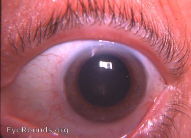
The patient was a 73-year-old white female with bilateral congenital mydriasis and no other abnormalities. The pupils reacted almost imperceptibly to light. Pharmacologic responses of the pupil to pilocarpine 4% solution, demecarium bromide 0.25%, and phenylephrine 10% indicated the presence of both the dilator muscle and the sphincter muscle but an abnormality relative to acetylcholine production in the iris.There was excellent response on the part of the pupil to 10% phenylephrine - indicating the presence of the dilator muscle.The family history of 8 female members with bilateral congenital mydriasis suggested an autosomal dominant inheritance mechanism with a possible degree of sex limitation.
Older patients usually have much smaller pupils than when they were young - an indication of decreased sympathetic neuro-effect on the dilator muscle.
Reference
Caccamise WC, Townes PL. Bilateral congenital mydriasis. Am J Ophthalmol. 1976 Apr;81(4):515-7. PubMed PMID: 944537.

Ophthalmic Atlas Images by EyeRounds.org, The University of Iowa are licensed under a Creative Commons Attribution-NonCommercial-NoDerivs 3.0 Unported License.