Focal scleral nodules (previously called solitary idiopathic choroiditis or unifocal helioid choroiditis) are rare, amelanotic, intrascleral lesions localized to the posterior segment of the eye. They are often elevated and yellow or white in color. These lesions are typically asymptomatic and are incidentally found on dilated fundus exam. It is uncertain whether the lesions are congenital or the result of an old inflammatory process. In the case shown here, there was overlying retinal pigment epithelium disruption without subretinal fluid. If asymptomatic, these lesions are observed without intervention.
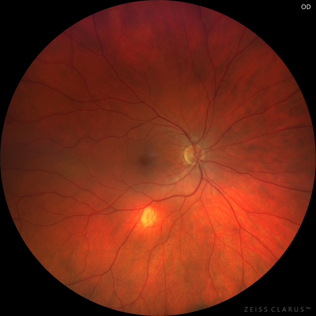
Figure 1: Fundus photography of the right eye showing a yellow-orange lesion inferior to the inferior arcade.
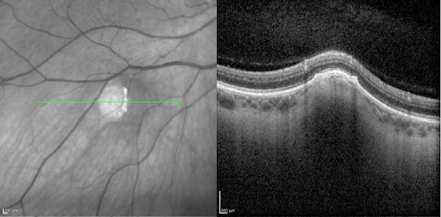
Figure 2: Dedicated OCT line scan over the lesion shows hyporeflective lesion in the sclera resulting in domed elevation of the overlying choroid and retina, without subretinal fluid.
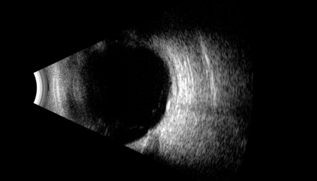
Figure 3: Longitudinal B-scan ocular ultrasound of the right eye at the 7 o'clock position demonstrating a small, localized calcified lesion inferotemporal to the nerve, corresponding to the focal scleral nodule.
University of Iowa
Roy J. and Lucille A. Carver College of Medicine
Department of Ophthalmology and Visual Sciences
200 Hawkins Drive
Iowa City, IA 52242
University of Iowa
Roy J. and Lucille A. Carver College of Medicine
Department of Ophthalmology and Visual Sciences
200 Hawkins Drive
Iowa City, IA 52242