This site uses tracking information. Visit our privacy policy. Click to agree to this policy and not see this again.

Ophthalmology and Visual Sciences

Bow-tie (band) atrophy secondary to compressive optic neuropathy

Bow-tie (band) atrophy secondary to compressive optic neuropathy

Category(ies): Neuro-ophthalmology
Contributor: Brittni A. Scruggs, MD, PhD; Matthew J. Thurtell, MD
Photographer: Susan J. Wright

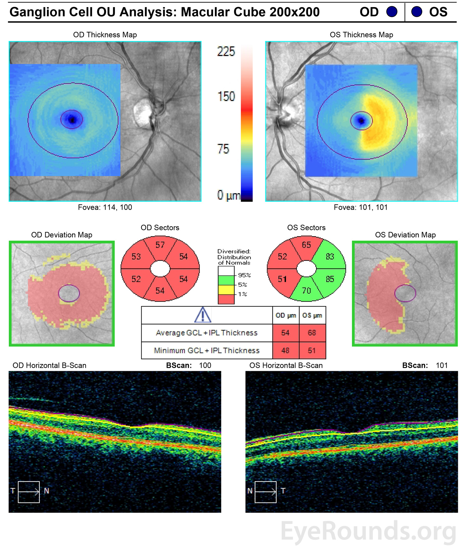
The patient was a 57-year-old man with vision loss in the right eye (OD) greater than left eye (OS) in the setting of a craniopharyngioma, which was resected 16 years prior. His visual acuity had been no light perception OD and 20/20 OS since the procedure.

Dilated funduscopic examination showed severe, diffuse optic atrophy OD and bow-tie (band) optic atrophy OS
Pupils were equal and reactive with a >3 log unit RAPD OD. Dilated funduscopic examination showed severe, diffuse optic atrophy OD and bow-tie (band) optic atrophy OS.
Goldmann visual fields showed a superior greater than inferior temporal defect OS
Goldmann visual fields showed a superior greater than inferior temporal defect OS.
vOcular coherence tomography of the macula showed diffuse ganglion cell layer (GCL) thinning OD and nasal GCL thinning OS
Ocular coherence tomography of the macula showed diffuse ganglion cell layer (GCL) thinning OD and nasal GCL thinning OS.
Summary:

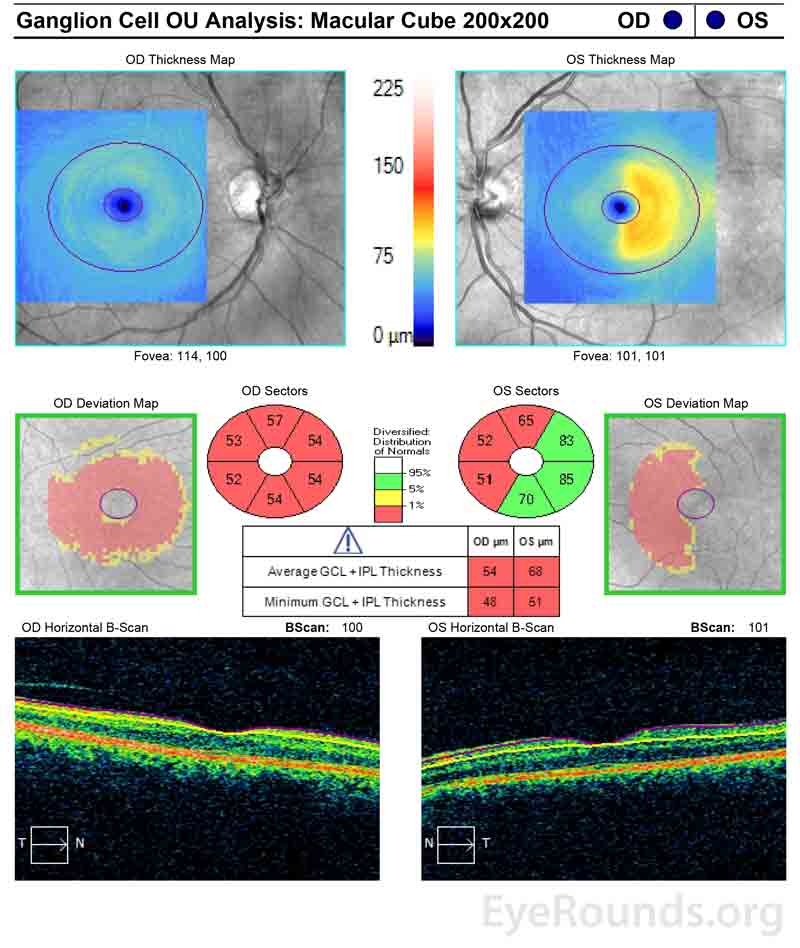
Contrast-enhanced magnetic resonance imaging (MRI) of the brain and orbits showed a stable tiny ring-enhancing cystic lesion anterior to the pituitary infundibulum and atrophy of the right greater than left optic nerves.

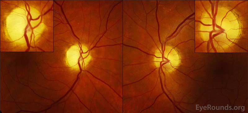
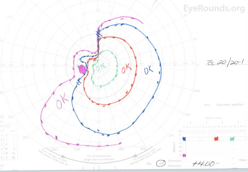
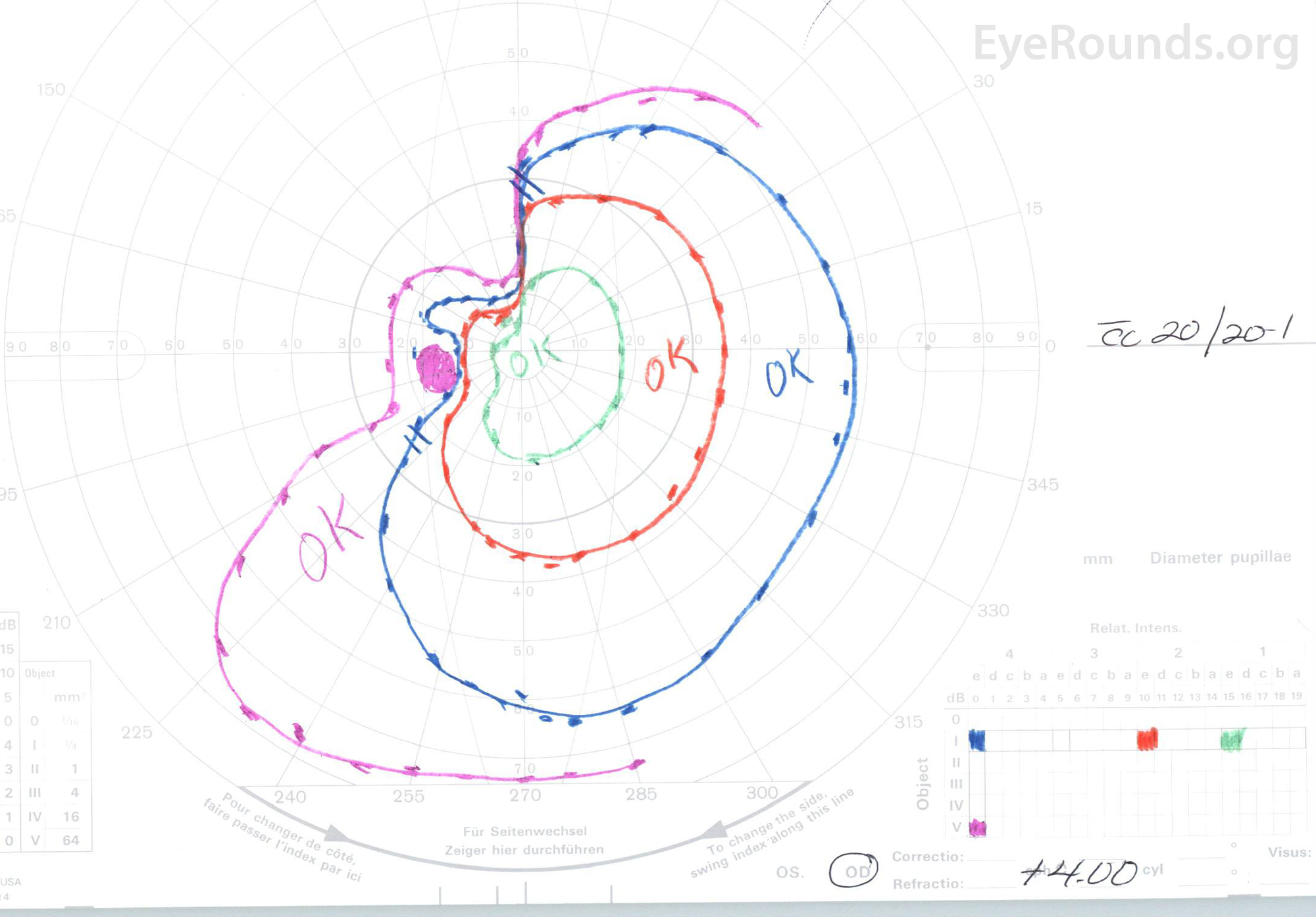
The presence of bow-tie, or band, atrophy suggests that there is a compressive optic neuropathy from an anterior chiasmal and/or medial, posterior optic nerve lesion. This clinical sign warrants further evaluation with magnetic resonance imaging (MRI) or computed tomography (CT) in a patient with unexplained vision loss. Unilateral band atrophy can also be seen in a patient with a unilateral optic tract lesion.