This site uses tracking information. Visit our privacy policy. Click to agree to this policy and not see this again.

Ophthalmology and Visual Sciences

Central Retinal Vein Occlusion

Central Retinal Vein Occlusion

Contributors: Sohan Singh Hayreh, MD, MS, PhD, DSc, FRCS, FRCOphth

Professor Emeritus of Ophthalmology
Ocular Vascular Clinic
Department of Ophthalmology and Visual Sciences
The University of Iowa
Roy J. and Lucille A. Carver College of Medicine


Introduction

Central retinal vein occlusion (CRVO) is a common cause of marked or total loss of vision in the middle-aged and elderly population, but no age group is immune to it. Although the disease entity has been known since 1878 and a large volume of literature has been published on the subject, its management is still ill-understood and controversial.

Since 1963 I have done experimental and clinical research on CRVO, its various types, course of the disease and various management options available and their merits and demerits. The following is a brief summary of my experience based on these studies and my way of managing CRVO


Types of Central Retinal Vein Occlusion

My studies showed that CRVO is actually of two types, with very different prognoses and management.[1-3] The two types are:

  • Non-ischemic type or venous stasis retinopathy.
  • Ischemic type or hemorrhagic retinopathy.

Classification of CRVO into non-ischemic and ischemic CRVO is essential because non-ischemic CRVO is a comparatively benign disease, with permanent central scotoma as the major complication from cystoid macular edema (see below). This type of CRVO does not develop the most dreaded complication of ocular neovascularization. In contrast to that, ischemic CRVO is a seriously blinding disease, and anterior segment neovascularization leading to neovascular glaucoma is its major complication (see below).


Relative Incidence of Ischemic Versus Non-Ischemic CRVO

In our series of 620 consecutive CRVO cases, we have found that 81% of the patients have the non-ischemic type, and only 19% are ischemic type[3]. That is the fortunate part of the condition: only one fifth of the patients suffer the serious type.

The first crucial step in the management of CRVO is to find out what type of CRVO an eye has because the prognosis, management and outcome of the two are totally different.


Criteria to Differentiate Ischemic from Non-Ischemic CRVO

Based on my experience of about 700 cases of CRVO over the years, I have developed diagnostic parameters to differentiate the 2 types of CRVO[1,2]. A prospective study of these parameters and their usefulness to differentiate ischemic CRVO from non-ischemic CRVO during the early phases of the disease revealed the following[2].

These tests can be divided into functional tests and morphological tests. Function tests consist of visual acuity, peripheral visual fields, relative afferent pupillary defect, and electroretinography. Morphological tests are: ophthalmoscopy and fluorescein fundus angiography.


Functional Tests

Visual Acuity: In our study we found that a visual acuity of better than 20/200 (6/60) was seen in 58% of the non-ischemic, as compared to only 1.7% in the ischemic type of CRVO. A visual acuity of better than 20/400 (6/120) was seen in 81% of the patients of the non-ischemic, as compared to about 7% of the ischemic type of CRVO only. A visual acuity of 20/400 or worse was seen in only 19% of the non-ischemic, whereas it was seen in 93% of the ischemic type of CRVO. Thus, our studies showed that if an eye with CRVO has 20/400 or worse vision, then there is about a 90% chance that that eye has ischemic CRVO.

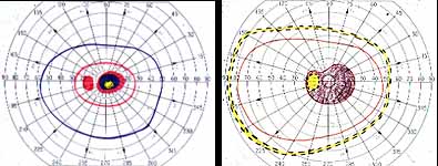
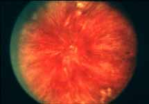
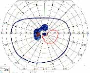
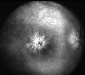
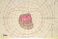
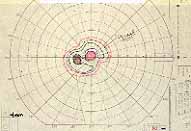
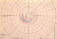
Peripheral Visual Fields: We find that peripheral visual fields, recorded with a Goldmann perimeter, give us very useful information in making this differentiation. Visual fields plotted with an automated perimeter do not help. We use, in the Goldmann perimeter, 3 isopters: I-2e, I-4e, and V-4e of the Goldmann perimeter. We find that in non-ischemic CRVO, the peripheral visual fields with V-4e and I-4e are perfectly normal, and they may even be normal with I-2e, or the I-2e is still seen in spite of the presence of peripheral visual field defects with this isopter. In ischemic CRVO, however, the eye may or may not see V-4e, or may or may not see I-4e, and usually does not see the I-2e target at all (Figure 1). Thus, in our study, in non-ischemic CRVO, all 3 targets were seen in 71% of the eyes, and I-4e and V-4e were seen in the remaining 29%, with no eye being unable to see any target or only V-4e. In contrast to that, in ischemic CRVO, only 8% of the eyes could see all the 3 targets, 63% saw I-4e and V-4e, and 18% only V-4e, and 10% could not see any target at all. Thus, if an eye can see I-2e or has normal visual fields with I-2e, that is definitely in favor of being a non-ischemic CRVO. If an eye can see only V-4e or no target at all, that eye definitely has ischemic CRVO.

visual fields
Figure 1. Visual Fields. Non-ischemic (left) and Ischemic (right) CRVO I-2e=Red I-4e=Blue III-4e=Brown V-4e=Yellow
Neutral density filters for quantifying afferent pupillary defect
Figure 2. Neutral density filters for quantifying afferent pupillary defect
RAPD in ischemic vs. non-ischemic CRVO
Figure 3. RAPD in ischemic vs. non-ischemic CRVO

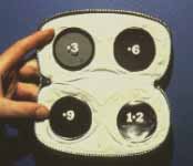
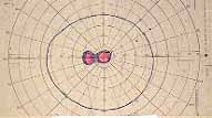
Relative Afferent Pupillary Defect (RAPD): The next test which helps us is the relative afferent pupillary defect. This is performed by a "swinging flashlight test". In a person with two normal eyes, if we shine the light on one eye, the pupil of that eye constricts immediately; then if we swing the light to the other eye, that pupil also constricts immediately. However, if one eye has retinal or optic nerve disorders, then if we first shine the light on the normal eye that pupil constricts, and then shine the light on the bad eye, instead of constricting the pupil dilates immediately in that eye; That is what is called a relative afferent pupillary defect. We can quantify the degree of relative afferent pupillary defect by using photographic neutral density filters (Figure 2). They come in 0.3, 0.6, 0.9 and 1.2 log units of transmission density, each 0.3 reducing the light by half. If we know that one eye has a relative afferent pupillary defect and the other eye is normal, then this filter is placed in front of the normal eye, not the bad eye. We start with 0.3 log unit filter in front of the normal eye. If on doing the test, the pupil in the bad eye still dilates, then we go to the next filter, 0.6 log unit. If it still dilates we go to the next filter, 0.9 log unit, and after that to the 1.2 log unit filter. (In fact, we can combine these filters to get higher log units.) So we keep doing that until the pupil in the bad eye starts to constrict instead of immediately dilating. That gives us the degree of the relative afferent pupillary defect.

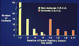
In our study we found that in non-ischemic CRVO, 97% of the eyes had a relative afferent pupillary defect of <0.6 log units. In contrast to that, in ischemic CRVO, 94% of the eyes had a relative afferent pupillary defect of >0.9 log units and 91% of the eyes >1.2 log units (Figure 3). Our studies also showed that all eyes with ocular neovascularization had relative afferent pupillary defects of >1.2 log units.

Electroretinography: We found that the electroretinography (ERG) parameter with the best sensitivity and specificity to differentiate ischemic from non-ischemic CRVO was amplitude of the b-wave (in both photopic and scotopic ERG). If the amplitude of b-wave is <60% of the normal, then there is an 80% chances that we are dealing with an ischemic CRVO. Or, if the amplitude of the b-wave is reduced by one or more standard deviation of the equipment, then we have a high chance of that being an ischemic CRVO. The advantage of ERG is that we can do this test even if the other eye is not perfectly normal, or in patients with only one eye. Relative afferent pupillary defect can only be tested when there is a perfectly normal second eye.


Morphological Tests

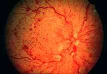
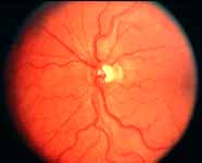
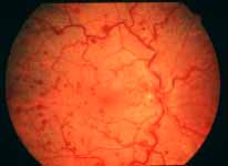
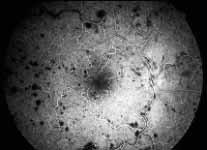
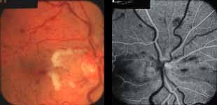
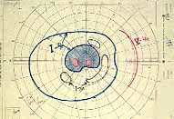
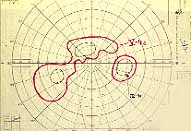
Ophthalmoscopy: Most ophthalmologists tend to use this as the main criterion to find out the type of CRVO. It is usually felt that extensive retinal hemorrhages and cotton-wool spots suggest an ischemic CRVO. However, our experience and our studies showed that these are very unreliable parameters. In fact our studies have shown that ophthalmoscopy is the least reliable and most misleading test for such a differentiation. For example, the eye in Figure 4 ophthalmoscopically looks like non-ischemic CRVO, but by all other parameters, it was an ischemic CRVO. Similarly, the fundus appearance of the eye in Figure 5a, with massive hemorrhages and few cotton-wool spots, is highly suggestive of ischemic CRVO; but the visual field plotting in Figure 5b shows that the eye can still see I-2e, and has normal peripheral visual fields with I-4e. As I mentioned earlier, if an eye can see I-2e there is a very high chance that that eye has non-ischemic CRVO. Moreover, this eye could see almost 20/40 - 20/50 which is another parameter telling us that this eye is a non-ischemic CRVO, in spite of extensive hemorrhages and some cotton-wool spots, which are considered signs of an ischemic CRVO. Two years later, the fundus and the angiographic appearances were perfectly normal in that eye - Figure 6a shows the fundus is perfectly normal, and Figure 6b shows fluorescein fundus angiogram with no capillary obliteration and perfectly normal retinal vascular beds; Thus, in fact this was a non-ischemic CRVO, which ophthalmoscopy would have made us believe was ischemic CRVO. Thus, our study has shown that the ophthalmoscopic appearances can be similar in ischemic and non-ischemic CRVO, during both early and late stages of the disease, and can mislead us in diagnosis.

Ophthalmoscopically looks like non-ischemic CRVO
Figure 4. Ophthalmoscopically looks like non-ischemic CRVO
fundus hemorrhages and few cotton-wool spots
Figure 5a. Massive hemorrhages and few cotton-wool spots
visual field example, Eye can still see I-2e
Figure 5b. Eye can still see I-2e, and has normal peripheral visual fields with I-4e
fundus example normal
Figure 6a. Fundus is perfectly normal
 fluorescein fundus angiogram with no capillary obliteration and perfectly normal retinal vascular beds
Figure 6b. Fluorescein fundus angiogram with no capillary obliteration and perfectly normal retinal vascular beds

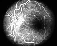
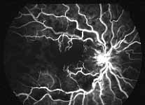
Fluorescein Fundus Angiography: In fluorescein fundus angiography, typically the retinal capillary non-perfusion or obliteration is considered the diagnostic criterion of ischemic CRVO. Figure 7a is a fundus photograph and Figure 7b an angiogram of an eye with non-ischemic CRVO, and typically we see that all the capillary bed fills very well, and there are a few scattered retinal hemorrhages and engorged retinal veins.

Fundus photograph of an eye with non-ischemic CRVO
Figure 7a. Fundus photograph of an eye with non-ischemic CRVO
An angiogram of an eye with non-ischemic CRV
Figure 7b. An angiogram of an eye with non-ischemic CRVO
Fundus almost total non-perfusion of the retinal capillary bed, indicating that this is an ischemic CRVO
Figure 8a. Fundus almost total non-perfusion of the retinal capillary bed, indicating that this is an ischemic CRVO
An angiogram almost total non-perfusion of the retinal capillary bed, indicating that this is an ischemic CRVO
Figure 8b. An angiogram almost total non-perfusion of the retinal capillary bed, indicating that this is an ischemic CRVO

In contrast to that, in Figure 8 there is almost total non-perfusion of the retinal capillary bed, indicating that this is an ischemic CRVO. If such good-quality angiogram of non-ischemic and ischemic CRVO is available, then fluorescein fundus angiography is very helpful. Unfortunately, there are multiple limitations in the evaluation of retinal capillary non-perfusion by fluorescein fundus angiography in CRVO, especially in fresh cases. These limitations include:

  • During the very early stages, in spite of retinal ischemia, retinal capillary non-perfusion may not be seen, because it takes time for the retinal capillaries to obliterate completely. For example, in Figure 9a the retinal capillary bed filled very well when the eye was first seen within a week or two after the onset of the visual loss, in spite of being an ischemic CRVO by other parameters. However, when seen 2 months later, the retinal capillary bed was completely obliterated (Figure 9b). So the first angiogram (Figure 9a) would make us believe that we were dealing with a non-ischemic CRVO.
  • If there are extensive retinal hemorrhages, as seen in Figure 10, it is usually almost impossible to evaluate capillary non-perfusion accurately, because of the masking effect by the blood.
  • Poor quality angiograms in these patients are not uncommon because of media opacity due to cataracts (because most of these patients are elderly), or a small pupil (because many of them have glaucoma and have been on Pilocarpine for a long time), or poor circulation (because of cardiovascular disorders), or poor cooperation, or other causes.
  • Inadequate sampling of the fundus is also a serious problem. We know that in diabetic retinopathy, CRVO and branch retinal vein occlusion, the earliest retinal capillary non-perfusion usually starts from the periphery and progresses toward the posterior pole. Conventional angiograms in these cases usually cover only the posterior pole, and miss extensive peripheral capillary non-perfusion. For example, in the eye shown in Figure 11a, the 60o angiogram of the posterior pole shows the capillary bed to be fairly good. However, the periphery of that eye showed extensive areas of capillary non-perfusion and even neovascularization (Figure 11b), so that an angiogram of the posterior pole here would point to non-ischemic CRVO, when, in fact, the eye has definite ischemic CRVO.

Thus, our study showed that fluorescein fundus angiography provided information on retinal capillary non-perfusion in only 2/3 of the eyes during the acute phase because of various limitations listed above. Not only that, but also fluorescein fundus angiography provided misleading information in some. Therefore, in our study, overall, angiography performed much worse than any of the functional tests. No doubt, if we can get good-quality retinal capillary information from all over the retina, fluorescein angiography is the best test.

Fallacy of using "10 disc area of retinal capillary obliteration" as the criterion to define ischemic CRVO: Another very important misleading factor in the various studies in the literature differentiating ischemic from non-ischemic CRVO is the widely advocated use of a "10 disc area of retinal capillary obliteration" as the normal yardstick. Our studies have shown that this is usually an invalid criterion for diagnosis of ischemic CRVO by fluorescein angiography. A recent multicenter study4 on CRVO came to the same conclusion. The study results clearly showed that eyes with less than 30 disc diameters of retinal capillary nonperfusion and no other risk factor are at low risk for developing iris/angle NV, "whereas eyes with 75 disc diameters or more are at highest risk". Thus "10 disc area of retinal capillary obliteration" is a poor and unreliable parameter in differentiating ischemic from non-ischemic CRVO as well as predicting ocular NV; and its use in many studies has resulted in misleading information and confusion.

retinal capillary bed filled very well
Figure 9a. The retinal capillary bed filled very well when the eye was first seen within a week or two after the onset of the visual loss, in spite of being an ischemic CRVO by other parameters.
fundus retinal capillary bed was completely obliterated
Figure 9b. 2 months later, the retinal capillary bed was completely obliterated
Extensive retinal hemorrhages<
Figure 10. Extensive retinal hemorrhages
fundus angiogram of the posterior pole shows the capillary bed to be fairly good
Figure 11a. 60o angiogram of the posterior pole shows the capillary bed to be fairly good
 capillary non-perfusion and even neovascularization
Figure 11b. The periphery of that eye showed extensive areas of capillary non-perfusion and even neovascularization
Fundus photographic comparison of ischemic and non-ischemic CRVO
Figure 12. Fundus photographic comparison of ischemic and non-ischemic CRVO

Conclusions Regarding Differentiation

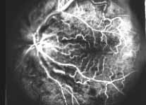
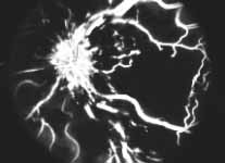
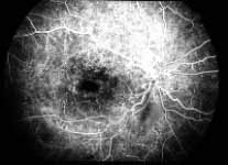
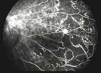
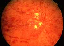
Table 1: The following table summarizes the sensitivities and specificity of the various functional tests.[2]

Functional tests

Sensitivity

Specificity

Visual acuity:

< 6/120

91%

88%

Peripheral visual fields (Goldmann):

No I-2e

97%

73%

Defective I-4e

92%

87%

RAPD:

> 0.9 log units

80%

97%

ERG:

b-wave amp.<60%

80%

80%

Based on our study[2] (Table 1), for differentiation of ischemic from non-ischemic CRVO during the acute phase, we found the overall order of reliability of these tests as follows:

  • Relative afferent pupillary defect is very reliable in uniocular CRVO, when the fellow eye is normal.
  • Next in order is ERG, which can be performed in all cases, even with bilateral CRVO or with only one eye.
    Combined information from the relevant afferent pupillary defect and ERG, provides the most reliable way of differentiating the two types of CRVO - in as many as 97% of the cases in our study.
  • Next in order of usefulness is the visual field plotting with a Goldmann perimeter, which is better than the visual acuity.
  • Angiography is usually much less useful than the functional tests during the acute stages of the disease. In fact, in our study, it provided definite information in only about 50% of the cases during the acute phase of the CRVO.
  • Ophthalmoscopy was the least reliable and the most misleading parameter. One cannot reliably diagnose whether the eye has ischemic or non-ischemic CRVO based purely on ophthalmoscopy (see fundus photographs in Figure 12).

A study without a clear distinction between the ischemic and the non-ischemic CRVO has little clinical or scientific validity, because, as I mentioned earlier, non-ischemic CRVO is a comparatively benign condition and there is no risk of neovascularization in this type of CRVO. In contrast to that, ischemic CRVO is a severely blinding disease, with a high incidence of anterior segment neovascularization, particularly, neovascular glaucoma.


Complications of CRVO

NON-ISCHEMIC CRVO: Long-term blinding complications are rare in this type. The major complication is chronic macular edema (Figure 13), leading on to cystoid macular degeneration, and permanent central scotoma; however, the peripheral visual field always remains normal. In about 12% of the eyes, within 18 months of onset non-ischemic CRVO may change to ischemic CRVO - more often in older than in younger persons[3]. If an eye with non-ischemic CRVO has a cilioretinal artery, then that eye develops cilioretinal artery occlusion (Figure 14) and an associated visual field defect and may produce sectoral optic atrophy.

Chronic macular edema
Figure 13. Chronic macular edema , leading on to cystoid macular degeneration, and permanent central scotoma
Cilioretinal artery occlusion
Figure 14. Cilioretinal artery occlusion

ISCHEMIC CRVO: There are many serious complications in this type. The most important complication is the development of ocular neovascularization, with neovascular glaucoma as the most dreaded complication. It is seen only in ischemic CRVO, and the overall incidence in our series was about 45% among the ischemic CRVO eyes. Other complications of ischemic CRVO are vitreous hemorrhage, macular degeneration, optic atrophy, retinitis proliferans, phthisis bulbi or loss of eye.

Ocular neovascularization[5]: As I mentioned earlier, 81% of the patients with CRVO have the non-ischemic type, and only 19% have the ischemic type, and ocular neovascularization attributable to CRVO does not occur in non-ischemic CRVO; There are, however two exceptions to this: i.e. eyes with associated (a) ocular ischemia and (b) background diabetic retinopathy, because some of these eyes, in spite of having non-ischemic CRVO, may develop ocular neovascularization. In ischemic CRVO the neovascularization is mainly of the anterior segment. Figure 15 is a graphic representation of cumulative chances of developing various types of neovascularization (NV) in ischemic CRVO.

graphs for or iris neovascularization, angle neovascularization, neovascular glaucoma, disc neovascularization and retinal neovascularization
Figure 15. On the X axis are the days from the onset of ischemic CRVO, and on the Y axis is the cumulative chance or probability of developing neovascularization. The various graphs are, from above down, for iris neovascularization, angle neovascularization, neovascular glaucoma, disc neovascularization and retinal neovascularization.

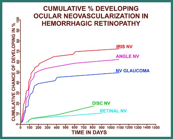
On the X axis for figure 15 are the days from the onset of ischemic CRVO, and on the Y axis is the cumulative chance or probability of developing neovascularization. The various graphs are, from above down, for iris neovascularization, angle neovascularization, neovascular glaucoma, disc neovascularization and retinal neovascularization. These graphs provide very important information on a number of issues, including the following:

  • Not every eye with ischemic CRVO develops ocular neovascularization because it depends upon the severity of retinal ischemia - more the ischemia, higher the chances of developing neovascularization.
  • When ocular neovascularization develops, the commonest site is the anterior segment, much less frequently the posterior segment.
  • The greatest risk of developing anterior segment neovascularization is during the first 7 months, after which the risk falls dramatically to minimal. This is extremely important information in the management of these cases; they need to be monitored very frequently during the first 7 months and if they do not develop neovascularization of the anterior segment during this period, then the risk of neovascular glaucoma drops to minimum.
  • The commonly held belief that every eye with CRVO that develops iris neovascularization or angle neovascularization is destined also to develop neovascular glaucoma is wholly wrong. In fact, about one-third of the eyes with iris neovascularization and about one-quarter of the eyes with both iris and angle neovascularization never develop neovascular glaucoma.

In order to place ocular neovascularization in overall CRVO in true perspective, it is important to point out two important facts: (i) Ischemic CRVO constitutes only one fifth of all CRVO cases. (ii) neovascular glaucoma, the most dreaded complication of CRVO, is seen at the maximum in about 45% of ischemic CRVO cases only. This would indicate that the overall incidence of neovascular glaucoma in the entire CRVO group is only 8 - 10% at the maximum - a highly important fact in any management considerations for CRVO.

Vitreous hemorrhage: In CRVO this may be either secondary to retinal/optic disc neovascularization or due to rupture of the blood through the internal limiting membrane, particularly in eyes with many sub-internal limiting membrane hemorrhages. In my experience, the latter source of vitreous hemorrhage is much more common than the former, and can occur in non-ischemic as well as ischemic types of CRVO, usually during the early stages of the disease. Vitreous hemorrhage from retinal/disc neovascularization usually occurs during the late stages of the disease. Sometimes the vitreous hemorrhage can occur from intraretinal microvascular abnormalities secondary to CRVO or development of posterior vitreous detachment. Therefore, it is important to be aware of the fact that the presence of vitreous hemorrhages in CRVO does not always mean the presence of retinal/disc neovascularization.


Natural History of CRVO

For proper management of a disease, understanding the natural history of the disease is absolutely essential, so that the natural history may not be interpreted as a beneficial effect of the treatment being advocated. I have discussed elsewhere the following aspects of natural history of CRVO.[6]

  1. Visual outcome: There is little definite information on the natural history of visual outcome in CRVO in the literature. The available information is based mostly on studies with either poor differentiation of ischemic from non-ischemic CRVO or none at all, most of them retrospective.[7,8,9] For example, the Multicenter CRVO study[7] reported that initial acuity largely determines the visual acuity outcome, but in that study CRVO was not differentiated into the two types. This invalidates the findings, because it is like mixing apples and oranges.
Table 2: Final visual acuity after resolution of retinopathy in my natural history study[10]

Visual acuity

Non-ischemic CRVO

20/15 to 20/40

65%

20/50 to 20/80

9%

20/100 to 20/200

11%

20/400

8%

CF or worse*

7%

Total eyes

144

* = Main cause of poor vision other than retinopathy, e.g., cataract, macular degeneration, glaucoma, etc.

CF = Counting fingers

  1. Ocular NV: As mentioned above, non-ischemic CRVO eyes do not develop NV unless there is associated disease that can cause NV, e.g., diabetes mellitus or ocular ischemia[5]. In ischemic CRVO: 
    • *the risk of developing anterior segment NV exists mainly during the first 7-8 months of the disease;
    • *the maximum risk of developing NVG is about 45%,
    • *and current conventional wisdom is wrong in assuming that all eyes with iris or angle NV go on to develop NVG; 1/3 of eyes with iris NV and ¼ of eyes with iris and angle NV do not progress to develop NVG on follow-up (Fig. 15). These three facts of the natural history of ocular NV in ischemic CRVO are of great clinical importance in its management.  
  2. Cumulative chances of conversion of non-ischemic CRVO to ischemic CRVO during follow-up: In our series of 500 eyes with non-ischemic CRVO, from the time of onset of non-ischemic CRVO, this happened within 6 months in 9.4% and reached almost its maximum within 18 months in 12.6% [3].  
  3. Resolution of retinopathy: In both types of CRVO, the retinopathy spontaneously resolves after a variable period. There is marked inter-individual variation in the time it takes to resolve - usually faster in younger than older people. Thus, both types of CRVO are self-limiting diseases, although during the period of activity they may produce various complications.

Management of CRVO

Management of CRVO still remains uncertain and highly controversial. Over the years many treatments have been advocated enthusiastically and success claimed but none has stood the test of time. These include anticoagulants, hemodilution, corticosteroids, acetazolamide, fibrinolytic agents, low molecular weight dextran infusion, carbon dioxide inhalation, vasodilators, hyperbaric oxygen, ocular hypotensive therapy, surgical decompression of CRVO, laser-induced chorioretinal anastomoses, photocoagulation, and a host of others.

The most important consideration when evaluating a therapy for any disease is to determine whether the therapy is based on incontrovertible scientific facts. Treatments without such a logical foundation prove not only useless but also sometimes harmful. Recently I have reviewed the various advocated modes of treatment in CRVO and their scientific validity [6] This review revealed that, unfortunately, most of the advocated treatments lack an incontrovertible scientific basis. Successes and beneficial effects claimed for many therapies in most cases simply represent the natural history of the disease, and that basic fact has been ignored in almost all the studies.

The fact that I would like to stress at the outset is that ischemic CRVO is a different, far more serious and potentially blinding disease than non-ischemic CRVO, and it requires wholly different management. Therefore, we must at first know which type of CRVO we are dealing with, and that makes differentiation of two types of CRVO the first essential step in the management of CRVO.

Following is a brief account of the major therapies advocated and tried from time to time. The main treatments can be divided into three categories: (A) medical, (B) surgical and (C) photocoagulation.

A. MEDICAL TREATMENTS

  • Anticoagulants and antiplatelet agents: The required treatment for many of the major systemic venous thromboembolic disorders (e.g., deep vein thrombosis) is anticoagulants or agents which reduce platelet aggregation. Because of that, unfortunately, there is a common impression among ophthalmologists and hematologists that these therapeutic agents should also help patients with CRVO. My studies of CRVO have shown anticoagulant therapy to be harmful because it increases the amount of retinal hemorrhages, with devastating results - often converting a benign form of CRVO into blinding CRVO[11]. Not only that, but I also have many patients who developed CRVO while on anticoagulants for various systemic diseases, clearly indicating lack of therapeutic benefit by anticoagulants. I have found that patients on antiplatelet agent, such as aspirin, are also liable to develop excessive retinal hemorrhages which adversely affect the outcome [11]

    Conclusion: Available evidence and our experience of managing more than 700 cases of CRVO over more than three decades indicate that anticoagulants and antiplatelet agents are contraindicated in CRVO[11]; they are not only of no therapeutic value but are definitely harmful. Therefore, they should never be used unless there are other more important life-threatening systemic indications.
  • Hemodilution: A few studies have suggested the presence of abnormal blood viscosity in CRVO patients. Based on that assumption, some authors have advocated the use of hemodilution in CRVO. I recently reviewed[6] all the available reports on hemodilution in the literature and found that there is little scientifically valid evidence of beneficial effects of this therapy.

    Conclusion
    : There is no convincing evidence that hemodilution benefits patients with CRVO. Therefore, I do not advocate this therapy.
  • Systemic corticosteroids: In non-ischemic CRVO, as discussed above, the primary cause of visual disability is the presence of macular edema. Therefore, in these cases the most important management consideration is controlling macular edema. There is no definite therapy available to treat macular edema. In our prospective studies on the subject for about 30 years, I have found that there is a small group of patients with non-ischemic CRVO who respond to systemic corticosteroids when given in high doses (80 mg Prednisone daily), with resolution of macular edema and visual improvement while they are on therapy. Unfortunately, when the steroids therapy is lowered below a certain level (usually 40 mg Prednisone), the macular edema returns and the vision starts to deteriorate. It is not at all uncommon for those who respond to treatment to require a maintenance dose of about 40 mg Prednisone or so for many months. For example, in one young man with non-ischemic CRVO in his only eye and a visual acuity of 20/200 (6/60) initially, visual acuity immediately improved to 20/30-20/40 (6/9-6/12) with 80 mg prednisone. To maintain that visual acuity he required a maintenance dose of 30-40 mg Prednisone for almost 3 years, and every attempt to go any lower immediately produced worsening of macular edema and deterioration of visual acuity. He finally ended up with a visual acuity of 20/20 (6/6) and no visual or systemic disability. Of course, a maintenance therapy for a long period produces side-effects of corticosteroids and the patient needs a close follow-up by both the ophthalmologist and the internist.

    The most critical thing to remember in systemic corticosteroid therapy for macular edema is that it does not work in every patient. If a patient shows no improvement within two weeks, I stop the treatment. It is also important to stress to the patient that the treatment is simply helping to reduce or eliminate macular edema to prevent long term permanent macular changes, and that it is NOT a cure for the CRVO which has to take its own natural course.
  • Intravitreal corticosteroids: Recently improvement of macular edema and visual acuity has been reported in a few eyes with non-ischemic CRVO and macular edema with intravitreal injection of triamcinolone acetonide. I have discussed this regimen of treatment in my recent review[6]. Intravitreal injection of triamcinolone acetonide temporarily improved visual acuity but on a long-term follow up there was no significant difference between the initial pre-injection visual acuity and the final visual acuity (personal communication from Professor Jost Jonas). Among the reported side-effects of intravitreal injection of triamcinolone acetonide (most of the studies are in diabetic macular edema) are development of ocular hypertension (requiring anti-glaucoma therapy) in about 33% to 50% of eyes after about one to two months, progression of cataract in some, and, rarely, endophthalmitis. Repeated intravitreal injections of triamcinolone acetonide can result in primary open angle glaucoma; this is particularly important in CRVO since in these patients there is already a significantly high incidence of glaucoma and ocular hypertension[12]. In contrast to this, in my experience of treating these patients with systemic steroids (see above), the incidence of developing ocular hypertension is very low.

    Conclusion
    : Available evidence indicates that oral or intravitreal corticosteroids may help to reduce macular edema associated with non-ischemic CRVO and improve visual acuity in some patients, but only as long as they are on the treatment. I have found steroid therapy of no help in ischemic CRVO in spite of the presence of macular edema, most probably because of irreversible ischemic damage to the retinal ganglion cell in the macular region.
  • Systemic acetazolamide (Diamox): I have found that some patients (but not all) with non-ischemic CRVO and macular edema respond to this therapy; but once again the macular edema is under control only so long as the patient is taking the drug. I usually give sustained release acetazolamide (Diamox Sequels) 500 mg twice daily. Unfortunately this drug can also produce severe systemic side-effects in some patients. If a patient does not respond within 2 weeks, there is high likelihood that he/she is not going to respond. I have not found this helpful in ischemic CRVO.

    Conclusion: Available evidence indicates that Diamox may help to reduce macular edema associated with non-ischemic CRVO and improve visual acuity for some patients, but only as long as they continue the treatment.
  • Ocular hypotensive therapy: Ophthalmologists often start the eye with CRVO on ocular hypotensive therapy, e.g., topical beta blockers, etc. or even systemic carbonic anhydrase inhibitors, under the erroneous impression that lowering the intraocular pressure in the involved eye improves the retinal blood flow. With venous outflow obstruction, lowering the intraocular pressure does not influence the retinal blood flow. Moreover, our clinical and experimental studies have clearly shown that development of CRVO, by some unknown mechanism, actually lowers the intraocular pressure, usually more than any of the ocular hypotensive agents do. We have discussed the whole subject in detail elsewhere[12]. I jokingly tell my residents and fellows that CRVO is the most effective treatment available to lower intraocular pressure! Thus, eyes with CRVO usually have normal intraocular pressure unless they develop neovascular glaucoma. In eyes with normal intraocular pressure, ocular hypotensive therapies seldom lower the pressure appreciably. Furthermore, lowering the IOP to very low levels could enhance macular edema.

    Since there is a high incidence of ocular hypertension or glaucoma in patients with CRVO, development of CRVO in one eye mandates treatment of ocular hypertension in the fellow uninvolved eye to reduce the risk of development of CRVO in that eye[12].

    Conclusion: There is no scientific rationale for the use of ocular hypotensive therapy in CRVO eyes with normal IOP. However, the fellow uninvolved eye MUST be treated to reduce the chances of its developing CRVO if it has ocular hypertension or glaucoma.

B. SURGICAL OR INVASIVE TREATMENTS

  • Fibrinolytic or thrombolytic agents: The theoretical objective of this therapy is to dissolve the preformed thrombus in the central retinal vein. From the studies reported in the literature there is little evidence that these have any beneficial effects [6]. Moreover, administration of these drugs can cause a significant hazard of bleeding, e.g., vitreous and cerebral hemorrhage and other systemic complications.

    A group[13] recently claimed enthusiastically that vitrectomy with branch retinal vein cannulation and infusion of t-PA in CRVO resulted in visual recovery which was "much better than what occurs as part of the natural history of CRVO". However, a critical review of that study reveals that the mode of treatment has no scientific rationale and that their claims are unwarranted. I have discussed the reasons for this at length elsewhere[14]. The claim of a result "much better than what occurs as part of the natural history of CRVO" was based on their comparison of visual acuity in their study with my natural history study findings [10] They stated: "In the study by Hayreh only 123 of 544 eyes with non-ischemic CRVO (23%) recovered vision to a level of at least 20/200". It mystifies me where they got their numbers of "123 of 544 eyes" in my paper[10] and how they came up with their interpretation. It is clear they misunderstood the visual acuity data in my paper (see Table 2 above)[10]. My natural history study[10] was based on 144 eyes with resolved non-ischemic CRVO (NOT 544 ) and it showed that in them the final visual acuity on resolution of CRVO was 20/40 or better in 65%, and it was better than 20/80 in 74%. When I challenged that misquotation [14], they offered a totally invalid, distorted interpretation of results of my study[15]. In fact, their entire response to the various issues raised in my comments had little logic[15]

    In addition, there is a considerable problem with the safety of the procedure. The authors claimed that it "is a relatively safe procedure" but their data shows that the extent of complications reported by the authors is so high as to be totally unacceptable[14].

    Conclusion: In spite of various claims made in various studies about the beneficial effect of thrombolytic therapy in CRVO, there is little scientifically valid evidence of its effectiveness for several reasons. This is particularly so since the thrombus organizes within few days and new capillaries and fibroblasts grow in from the vessel wall to convert it into vascular connective tissue which cannot be dissolved by thrombolytic agents given by any route by the time most CRVO patients come for consultation. Moreover, thrombolytic therapy has serious ocular and even fatal systemic side-effects.
  • Surgical decompression of CRV: I have recently discussed in detail the problems and dangers of this surgical procedure[6] This procedures is of NO scientific merit at all, and could be dangerous. Of particular concern in this context is the recent claim by Opremcak et al.[16] about surgical decompression of the CRVO by a procedure they called "radial optic neurotomy". They made a radial cut from the vitreous side in the optic nerve head, extending all the way down to the lamina cribrosa, adjacent sclera and cutting the arterial circle of Zinn and Haller. They claimed that "radial optic neurotomy" is a "surgically feasible and safe procedure" and is "beneficial". I have researched various basic and clinical aspects of the optic nerve head and CRVO since 1955[17]. Based on that, I find that this procedure lacks any scientific rationale, could be harmful, and comes without reliable evidence that it has any beneficial effect. I have discussed at length elsewhere the various problems with this procedure and their claims[17].

    Conclusion: The whole idea of surgical decompression of central retinal vein in CRVO is ill-conceived and based on lack of basic understanding of: (a) the anatomy of the optic nerve and CRV and their relationship with each other, (b) the site of thrombosis in central retinal vein, and (c) the fact that a vein completely closed by a thrombus cannot be opened by "decompression" because the thrombus organizes shortly after it is formed (see above). Above all, it is a dangerous procedure.
  • Laser-induced chorioretinal venous anastomosis for treatment of non-ischemic CRVO: This is one of the treatment modalities being advocated for nonischemic CRVO. To evaluate the role of these iatrogenic chorioretinal anastomoses in the management of CRVO, let us review the available information on it. McAllister and Constable[18], in 1995 first reported producing chorioretinal anastomoses experimentally in eyes with nonischemic CRVO by using high power density argon laser photocoagulation to one of the retinal veins in the lower part of the fundus at least 3 disc diameter away form the optic disc. Based on their studies, they advocated laser-induced chorioretinal anastomosis as a treatment modality for nonischemic CRVO. In order to judge the appropriateness of this mode of treatment, one has to evaluate its benefits and risks from the available data.
     
  •  Visual outcome: There are only two series with any appreciable number of cases reported so far. McAllister and Constable18 in their original study in eyes with nonischemic CRVO, were able to produce successful chorioretinal anastomoses in only 8 of 24 eyes (33%) in 3 to 7 weeks, after 1 to 5 laser attempts. Among the 8 eyes with successful anastomosis, 6 eyes with initial visual acuity of about 20/200 had final visual acuity of 20/200 in 2, 20/120 in one, 20/60 in 2, 20/20 in one; and 2 eyes with initial visual acuity of 20/120 improved to 20/30. Fekrat et al.[19], successfully produced the anastomosis in 9 of 24 (38%) eyes with nonischemic CRVO, within 8 weeks after the laser application. Six of the 9 eyes in this series already had developed retinociliary collaterals on the optic disc before the laser procedure was performed so that nature had already performed what this iatrogenic procedure was meant to do. Browning and Antoszyk[20] tried laser-induced anastomosis in 8 eyes with nonischemic CRVO (successful anastomosis developed in only 2 eyes), and reported visual acuity improvement in 2 independent of failed attempts at anastomosis creation and did not improve or worsened in 6 eyes, including the 2 with the successfully created anastomosis. The conventional "two or more line" improvement in visual acuity occurred in 5 of 8 (62.5%) eyes of McAllister and Constable[18] and in 3 of 9 (33%) eyes of Fekrat et al.[19], with no significant improvement in the rest. The authors of these two latter series claimed that none of the eyes with successful anastomosis progressed to ischemic CRVO. 

Complications: Various studies have reported a number of complications which can be divided into immediate and late complications.

    1. Immediate complications: Among the immediate complications noticed at the time of laser application, those reported by McAllister and Constable[18] included hemorrhages from the retinal vein (in 40%), subretinal hemorrhages (in 7%), and choroidal hemorrhages (in 5%). Fekrat et al.[19] reported transient vitreous hemorrhage at the time of laser application in 42%. In my studies, producing experimental branch retinal vein occlusion by argon laser application in rhesus monkeys in the 1970s, I found that retinal, subretinal, choroidal and vitreous hemorrhages commonly developed at the time of laser application.
    2. Late complications: McAllister and Constable[18] reported branch retinal vein occlusion with segmental retinal ischemia (in 11%), preretinal (in 13%) and subretinal (in 5%) fibrosis at the site of the laser application, and development of preretinal neovascular frond over the site of laser application. Fekrat et al.[19] reported localized choroidal neovascularization in 21%, mild preretinal fibrosis in 8%, and preretinal fibrosis and traction retinal detachment requiring vitrectomy in 4%. However, the authors claimed that there were no permanent vision-limiting complications. By contrast, Browning and Antoszyke[20] attempted to produce laser-induced venous anastomosis in 8 eyes with nonischemic CRVO (with 20 attempts they had 2 successful anastomoses but not of a therapeutic type), and of those 8 eyes, 3 developed iris neovascularization, retinal neovascularization at the laser site and vitreous hemorrhage, 2 developed traction retinal detachment, and one neovascular glaucoma. These eyes required secondary panretinal photocoagulation (in 3), pars plana vitrectomy (in one) and glaucoma seton implant (in one). They concluded that laser-induced chorioretinal anastomosis for nonischemic CRVO has greater risk and less success than the initial reports suggested, and that successful chorioretinal anastomosis does not preclude development of anterior segment neovascularization. In addition to these reports, during 1996-98, there are 4 more published reports of serious complications with this procedure; the reported complications include choroidal neovascular frond into the vitreous followed by vitreous hemorrhage, recurrent vitreous hemorrhage, deep subretinal hemorrhage, subretinal choroidal neovascular membrane, massive preretinal fibrosis, traction retinal detachment, and neovascular glaucoma. All these late complications were reported usually within the first few months after the procedure. The possibility of still more long-term complications cannot be ruled out.

To place the value of laser-induced chorioretinal venous anastomosis for treatment of nonischemic CRVO in true perspective, one has to consider the natural history of nonischemic CRVO (see "Natural history of CRVO" above) .

When evaluating the validity of claims of improved visual acuity in any study in the literature, one has to be aware of the possibility that apparent "improvement" in visual acuity can simply be the result of multiple artifacts in visual acuity testing (which is often done by technicians). In my experience of repeated testing of visual acuity, myself, in about three thousand patients with various circulatory disorders of the eye (e.g., anterior ischemic optic neuropathy, central and branch retinal artery occlusion and central and branch retinal vein occlusion, ocular ischemia) over time, I have found that the most important factor in apparent "improvement" of visual acuity may simply be a patient's improved skill in reading the visual acuity chart; he/she may have learned by experience to read the test chart better by looking around and fixating eccentrically. This applies particularly to an eye which has a visual field defect or a scotoma passing through or just involving the central fixation spot. (Eyes with nonischemic CRVO invariably have poor visual acuity due to central scotoma.) That is why reports of improved visual acuity without corresponding improvement of central visual field defects can be misleading.21 None of the studies reported concurrent improvement of visual acuity and visual field.

It is therefore evident from the available data that the visual outcome apparently achieved in eyes with nonischemic CRVO by laser-induced chorioretinal venous anastomosis seems no better, if not worse, than the natural history of the disease. On the top of that, the procedure is associated with a fairly high risk of serious vision threatening complications, not seen in non-ischemic CRVO

Conclusion: It is evident that the complications of laser-induced chorioretinal venous anastomosis for treatment of non-ischemic CRVO heavily outweigh any dubious benefits, and that this is not a safe and effective mode of treatment for a condition which has a fairly good outcome if simply left alone (see "Natural history of CRVO" above).

C. PHOTOCOAGULATION TREATMENT

In the management of CRVO, panretinal photocoagulation has been widely advocated as the treatment of choice. The presumed role of panretinal photocoagulation in retinal diseases is to prevent development of neovascularization in eyes with retinal ischemia.

Non-ischemic CRVO: Since there is no retinal ischemia in this type of CRVO, and thereby no risk of developing neovascularization[5], there is no scientific basis at all for doing panretinal photocoagulation in non-ischemic CRVO. Moreover, panretinal photocoagulation is not a harmless procedure; it can produce loss of peripheral visual fields in CRVO (see below - Figures 16,17) and may cause defective vision in the dark. Some ophthalmologists advocate the use of macular grid photocoagulation for microcystic macular edema; however, a recent multicenter CRVO photocoagulation study revealed that it has no beneficial effects in these eyes and does not improve visual acuity[22]. In my experience, some patients may land up with much worse central scotoma from such a treatment. Thus, photocoagulation has no role whatsoever in the management of non-ischemic CRVO.

Ischemic CRVO: It has been almost universally accepted that prophylactic panretinal photocoagulation is the treatment of choice to prevent neovascular glaucoma or treat neovascular glaucoma itself. I have found little definite scientific proof in support of this assumption[23]. Therefore, we investigated this in a 10-year prospective study, to find out its effect on ocular neovascularization, particularly neovascular glaucoma associated with ischemic CRVO. The results, which were very surprising, were published in 1990[23]. In summary, our study showed that, on comparing the lasered eyes versus the non-lasered eyes, there was no statistically significant difference between the two groups in the incidence of development of: (a) angle neovascularization, (b) neovascular glaucoma, (c) retinal and/or optic disc neovascularization, and (d) vitreous hemorrhage, and (e) in visual acuity. Our study did show a statistically significant (p=0.04) difference in the incidence of iris neovascularization between the two groups, with iris neovascularization less prevalent in the lasered group than in the non-lasered group, but only when the laser was performed within 90 days after the onset of CRVO; however, iris neovascularization per se produces no harmful effect in the eye and as such is of little importance. The most important result of this study was the markedly deleterious effect of panretinal photocoagulation on the peripheral visual fields, because the lasered group suffered a significantly (p<0.03) greater loss than the non-lasered group (Figures 16, 17). Thus, contrary to the prevalent impression, our study showed that panretinal photocoagulation has no statistically significant beneficial effect in reducing neovascular glaucoma. On the other hand, it markedly damages the peripheral visual field, and may convert an eye with normal peripheral vision into almost a blind eye.

Visual Field
Figure 16. I-4e=Blue III-4e=Brown V-4e=Red

see also Figure 17. Visual Field Examples before and after Pan Retinal Photocoagulation Recently, a multicenter clinical trial by "the Central Vein Occlusion Study" (CVOS) group investigated the role of panretinal photocoagulation in ischemic CRVO.4 The purpose of that study was twofold: (1) to find out whether prophylactic panretinal photocoagulation prevents development of iris and angle neovascularization, and (2) whether panretinal photocoagulation prevents progression of iris/angle neovascularization to neovascular glaucoma.

  1. In answer to the first question, the study reported that "prophylactic panretinal photocoagulation does not totally prevent" development of iris/angle neovascularization and concluded that prompt regression of iris and angle neovascularization in response to panretinal photocoagulation is more likely to occur in eyes that have not been treated previously prophylactically. This study should lay to rest the claims that prophylactic panretinal photocoagulation in ischemic CRVO prevents the development of iris and angle neovascularization.
  2. As for the second aspect of the study, the authors recommended "careful observation with frequent follow-up examinations in the early months (including undilated slit-lamp examination of the iris and gonioscopy) and prompt panretinal photocoagulation of eyes in which 2'clock iris/angle neovascularization develops." I have serious reservations regarding the validity of the findings in the CVOS, as to whether panretinal photocoagulation in ischemic CRVO actually prevents the development of neovascular glaucoma. Such a multicenter, multimillion dollar study conducted under the aegis of the National Institute of Health carries tremendous prestige and its conclusions become a weighty verdict. Thus, it is vital to discuss this aspect of the study at some length, to place its results in their true perspective for the ophthalmic community. I have discussed that elsewhere[24].

The most important feature of any study is its design, because that can determine its conclusions and their validity. Based on my clinical and experimental study on CRVO during the past 30 years, I have some important concerns about the baseline design of the study[25]. It is now well-accepted that neovascular glaucoma is a complication only of ischemic CRVO and is NOT seen in non-ischemic CRVO. For a study claiming beneficial effects of panretinal photocoagulation on anterior segment neovascularization in ischemic CRVO, it is imperative to ask at least the following three very basic questions:

  1. Did all the patients in the study have ischemic CRVO?
  2. Were the results of panretinal photocoagulation therapy better than the natural history of anterior segment neovascularization in untreated ischemic CRVO?
  3. What are the side-effects and complications of the therapy?
  • Did all the patients in the study have ischemic CRVO? The first logical step in conducting such a study is to differentiate ischemic from non-ischemic CRVO accurately. The design used in the study to differentiate the two types of CRVO at the baseline entry level has serious problems, including the following:
    • The investigators used "10 disc area of retinal capillary nonperfusion" on fluorescein angiography as almost the sole criterion for differentiation between ischemic and non-ischemic CRVO. There are many pitfalls in using this criterion. All the available evidence indicates that the more marked the retinal ischemia, the earlier and more frequent is the ocular NV[5]. Anterior segment neovascularization in ischemic CRVO depends upon the global retinal ischemia[5]. The CVOS also showed that iris/angle neovascularization correlated with the amount of nonperfused retina (p=0.0001)[4]. The study itself proved that their criterion of 10 disc area retinal capillary nonperfusion was not an appropriate one to differentiate the two types of CRVO; they found that eyes with less than 30 disc diameters of nonperfusion and no other risk factor are at low risk for iris/angle neovascularization, "whereas eyes with 75 disc diameters or more (i.e., eyes that show virtually no intact capillaries in the posterior pole) are at highest risk"[4]. This has also been our experience. Also, "eight standard views" by 30o fundus photography (utilized in a proportion of their cases) do not usually outline the peripheral retinal vascular bed satisfactorily, and therefore do not reveal the changes in the entire peripheral part of the retinal vascular bed - which is frequently the first area to develop capillary nonperfusion[2,5]. Moreover, the "10 disc area of retinal capillary nonperfusion" may simply represent focal retinal ischemia seen in some non-ischemic CRVO.

      In contrast to the criterion of "10 disc area of retinal capillary nonperfusion" , our study[2] showed that, during the acute phase, the information provided by four visual function tests (i.e. visual acuity, kinetic perimetry, RAPD and ERG), taken together, is far superior, not only in separating the ischemic from non-ischemic CRVO more effectively and reliably than fluorescein angiography alone, but also in giving good information about the extent of global retinal ischemia (as discussed above). Thus, fluorescein angiography is a weak staff to lean upon in making such a differentiation during the early acute phase of CRVO, because angiography at this stage may provide no information or misleading information on retinal capillary non-perfusion in at least one third of patients (see above)[2]. As is evident from the above discussion, this was also proved by the CVOS.
    • There is also other evidence in the CVOS baseline data[25] suggesting that some of their non-ischemic CRVO eyes probably had ischemic CRVO initially, and vice versa. For example:
      • In the CVOS baseline information, 21% of the eyes classified as having ischemic CRVO had a visual acuity of 20/20 to 20/100[25] - we found that in only 1.7% of eyes with ischemic CRVO[2].
      • In their eyes where panretinal photocoagulation was not done until they developed iris/angle neovascularization, the neovascularization developed in 35% of the eyes overall, during the study period[4]. As discussed above, we investigated the same in our natural history study on ocular neovascularization in ischemic CRVO, and found that 70% of ischemic CRVO eyes develop iris NV[5] (Figure 15) - double the incidence seen in the ischemic CRVO cases in CVOS. This, again, suggests that some of the 91 eyes in the CVOS group did not have ischemic CRVO.
      • In the CVOS baseline data[25], at 4-month follow-up at least 16% of the non-ischemic CRVO eyes "developed evidence of ischemia" (i.e. converted to ischemic CRVO)[25] - this is a far higher incidence than that reported in the literature[26] and seen by us[3]. In our 500 eyes with non-ischemic CRVO studied prospectively, we found the cumulative chance of conversion only 8-9% at 6 months after the onset[3]. This suggests again that some of their "non-ischemic CRVO" eyes probably had ischemic CRVO to begin with. As discussed above, this may have been caused by a fluorescein angiographic artifact, because during the very early stages of ischemic CRVO, in spite of retinal ischemia, angiography may show minimal retinal capillary non-perfusion; we have found that the extent and severity of retinal capillary nonperfusion in ischemic CRVO tends to increase with time[2,5].

        From the above discussion, it is evident that the criterion of "10 disc area of retinal capillary obliteration" is a poor and unreliable parameter for differentiating ischemic from non-ischemic CRVO, as well as for predicting ocular neovascularization. Thus, their baseline data[25] suggest that they had a mixture of both ischemic and non-ischemic CRVOs in their two types of CRVO - such a mixture has the potential of giving misleading information.
  • Are the results of panretinal photocoagulation therapy better than the natural history of anterior segment neovascularization in untreated ischemic CRVO? In judging the outcome of any therapy, the first and most important essential is to know the natural history of the disease. The authors of the CVOS assumed that any ischemic CRVO eye with 2 clock hours of iris/angle neovascularization is certain to go on to develop neovascular glaucoma and therefore deserves prompt mandatory panretinal photocoagulation.4 But this is a false assumption and ignores the natural history of iris and angle neovascularization. We studied this important aspect in our prospective natural history study on ocular neovascularization in ischemic CRVO[5]. Our findings are shown in figure 15. About one third of the eyes with iris neovascularization and about one quarter with iris/angle neovascularization never developed neovascular glaucoma on follow-up[5]. Our criterion of neovascular glaucoma was persistent elevated IOP above 21 mmHg. Iris neovascularization may be worrisome and an indication for closer observation but I have followed some ischemic CRVO eyes with iris/angle neovascularization very closely for years, and found they never progressed to develop neovascular glaucoma, and the iris and angle neovascularization resolved spontaneously. The primary objective of panretinal photocoagulation is to prevent development of neovascular glaucoma.In my studies iris/angle neovascularization on their own have no long-term deleterious complications - it is only if such an eye develops neovascular glaucoma that the eye suffers damage. Much more importantly, by treating all patients with iris neovascularization, and not randomizing them to "treatment" or "no treatment", the study left a serious cloud over its use of iris neovascularization as an outcome measure for development of neovascular glaucoma, because about one third of the eyes treated with panretinal photocoagulation, which would never have developed neovascular glaucoma (Figure 15), were unnecessarily subjected to the risk of developing marked and crippling peripheral visual field loss (as discussed above)[23]. In our study on argon laser panretinal photocoagulation in ischemic CRVO[23], although eyes with panretinal photocoagulation showed a significantly (p=0.04) less prevalent iris neovascularization in the panretinal photocoagulation group than in the control group, there was no statistically significant difference between the two groups in angle neovascularization and neovascular glaucoma.
  • What are the side-effects and complications of the therapy? Most importantly, no consideration was given in the CVOS design to obtaining information on the effect of panretinal photocoagulation on peripheral visual fields. Eyes with ischemic CRVO almost always have a large permanent central scotoma, resulting in poor central visual acuity. Like our study[23], the CVOS[4] also showed no beneficial effect from panretinal photocoagulation on visual acuity. In spite of a large central scotoma, untreated eyes usually retain good "getting around" peripheral vision because their peripheral fields are preserved (similar to age-related macular degeneration), if the eye does not develop uncontrolled neovascular glaucoma (see visual fields before panretinal photocoagulation in Figures 16, 17). But in our study[23], as discussed above, we found a statistically significant (p<0.03) worsening of peripheral visual fields, with marked loss, in eyes treated with panretinal photocoagulation as compared to those in the control non-laser group; figures 16 and 17 includes some examples of that. Following panretinal photocoagulation, the large central scotoma combined with severe loss of peripheral visual fields may virtually blind the eye. Should we destroy most of an eye's remaining useful peripheral vision with panretinal photocoagulation unless we are quite certain that, without treatment, every eye with iris and angle neovascularization is destined for a painful death? This study does not provide justification for that.

    A study with such flaws in its basic design has the potential to provide misinformation which may retard rather than advance knowledge, with regard to the role of panretinal photocoagulation in ischemic CRVO. Although our study[23] and that reported by CVOS[4] both deal with the role of argon laser panretinal photocoagulation in ischemic CRVO, because of the difference in the basic designs of the two studies, they are very different.

    It could very well be argued that this is simply my opinion, which might not necessarily be accepted by the authors of the study. As is customary, the Editor of Ophthalmology (where this study was published) sent my comments to the authors of the CVOS Group for their comments. Following was their response[27]: "Dr. Hayreh raises a number of excellent points, based on his extensive research and clinical studies over the past decades. .…. We agree with Dr. Hayreh that our minimum criteria of 10 disc areas of retinal capillary nonperfusion for defining ischemic vein occlusion is a low risk for development of iris neovascularization (INV)…. We also agree with Dr. Hayreh that when there is too much intraretinal hemorrhage to evaluate perfusion on the fluorescein angiogram, such eyes are likely to be nonperfused (ischemic CRVO). ……We agree that INV never develops in many eyes with ischemic vein occlusion. ….. Dr. Hayreh is correct that we were not willing to follow the natural history of eyes in which INV was developing because of our fear that neovascular glaucoma would develop quickly; consequently, we do not have information about the natural history after INV develops in untreated eyes."
  • Difference in response to panretinal photocoagulation of ischemic CRVO compared with proliferative diabetic retinopathy: Another important question that needs to be answered here: if panretinal photocoagulation helps to reduce the incidence of neovascular glaucoma in proliferative diabetic retinopathy, why it does not do the same in ischemic CRVO? But the two disease processes are entirely different. The original rationale for advocating panretinal photocoagulation in ischemic CRVO was the proven beneficial effect of panretinal photocoagulation on ocular neovascularization in proliferative diabetic retinopathy; however, this did not take into consideration the marked disparities in the disease processes between ischemic CRVO and proliferative diabetic retinopathy and in their responses to panretinal photocoagulation. Proliferative diabetic retinopathy is a chronic, slowly progressive disease in a person with an incurable basic systemic disease. By contrast, ischemic CRVO is an acute catastrophe associated with severe and extensive retinal ischemia but with a self-limiting course. One can compare ischemic CRVO to a hurricane which develops suddenly, lasts for a short period but inflicts extensive, devastating damage to a house, whereas diabetic retinopathy is like a slow leak which gradually undermines the house over the years, slowly and insidiously. Measures which would successfully control the damage caused by a slow leak are useless against a hurricane! The extent of retinal ischemia, and hence the quantity of the presumed vasoproliferative factor(s), in ischemic CRVO is many, many times that in average proliferative diabetic retinopathy. Panretinal photocoagulation may be able to cope with the mild amount of retinal ischemia seen in the usual proliferative diabetic retinopathy but is totally inadequate and ineffective when there is the severe, extensive, sudden retinal ischemia of ischemic CRVO. This basic fact has been totally ignored by the advocates of panretinal photocoagulation.

When all these facts are put together, a very different perspective on ischemic CRVO emerges - and, consequently, a different perspective on its management. In ischemic CRVO, if the eyes either do not develop neovascular glaucoma (and at least 50% will not - Figure 15) or their high IOP due to neovascular glaucoma is controlled satisfactorily by the means discussed below, once the retinopathy burns itself out, they are frequently left with a large, dense, absolute central scotoma (from permanent macular damage invariably seen in this disease), but with a normal or a reasonably good peripheral visual field. The latter is very useful for "getting around" in daily life. By contrast, in ischemic CRVO eyes treated with panretinal photocoagulation, the eye is left with extremely limited visual capabilities, due to a combination of large absolute central scotoma with marked visual field constriction. Therefore, panretinal photocoagulation does more harm than good to the majority of these eyes (Figures 16,17); it neither confers a statistically significant protection against neovascular glaucoma, nor always prevents development of ocular neovascularization, nor confers any other significant benefit. This is particularly undesirable in the 50% or more of ischemic CRVO eyes which would never have developed neovascular glaucoma anyway (Figure 15) and thus did not need panretinal photocoagulation. The high risk of marked peripheral visual field loss from panretinal photocoagulation combined with the naturally developing central scotoma from the disease itself (Figures 16,17), is a serious disabling complication of panretinal photocoagulation without any countervailing benefit.

Conclusion of role of panretinal photocoagulation in ischemic CRVO

As is evident from the discussion above, there is no scientifically valid proof so far that panretinal photocoagulation is safe and effective in prevention or management of neovascular glaucoma in acute ischemic CRVO. In spite of that and ignoring the fact that panretinal photocoagulation is highly destructive to the remaining peripheral visual fields in most of the acute ischemic CRVO cases (Figures 16,17), there are authors who still actively advocate its use[4,28].


Our Management Regimen for Ischemic CRVO

Since neovascular glaucoma is the most dreaded complication of ischemic CRVO, naturally the question arises how to manage ischemic CRVO. For a logical management of any disease, first one has to understand the basic issues involved and the information available which should act as guidelines. In ischemic CRVO, we have currently the following information available:

graphs for or iris neovascularization, angle neovascularization, neovascular glaucoma, disc neovascularization and retinal neovascularization
Figure 15 repeat. On the X axis are the days from the onset of ischemic CRVO, and on the Y axis is the cumulative chance or probability of developing neovascularization. The various graphs are, from above down, for iris neovascularization, angle neovascularization, neovascular glaucoma, disc neovascularization and retinal neovascularization.
  • A maximum of about 45% of ischemic CRVO patients are likely to develop neovascular glaucoma (Figure 15), (contradicting the common impression among ophthalmologists that a vast majority of these eyes develop neovascular glaucoma); 55% are never going to develop it[5].
  • The risk of developing neovascular glaucoma is mainly during the first 7-8 months of the disease (about 40%)[5] (Figure 15). After that the risk falls dramatically to 5% or less. So the most crucial period to monitor these patients closely is first 7-8 months.
  • The multi-center CRVO study Group showed that prophylactic panretinal photocoagulation in ischemic CRVO does not prevent iris and angle neovascularization[4]. 
  • Our panretinal photocoagulation study showed that eyes subjected to panretinal photocoagulation usually suffer marked loss of peripheral visual fields[23] (Figures 16,17). Combined with the large central scotoma in these eyes, that peripheral visual fields loss can make these eyes almost blind.
Visual Field
Figure 16. I-4e=Blue III-4e=Brown V-4e=Red
Visual Field
Figure 17a. Before PRP,-4e=Blue III-4e=Brown V-4e=Red
Visual Field
Figure 17b. After PRP, -4e=Blue III-4e=Brown V-4e=Red
Visual Field
Figure 17c. Before PRP,-4e=Blue III-4e=Brown V-4e=Red
Visual Field
Figure 17d. After PRP, -4e=Blue III-4e=Brown V-4e=Red
Visual Field
Figure 17e. Before PRP,-4e=Blue III-4e=Brown V-4e=Red
Visual Field
Figure 17f. After PRP, -4e=Blue III-4e=Brown V-4e=Red
Visual Field
Figure 17g. Before PRP,-4e=Blue III-4e=Brown V-4e=Red
Visual Field
Figure 17h. After PRP, -4e=Blue III-4e=Brown V-4e=Red

I find no convincing scientific evidence that panretinal photocoagulation usually helps prevent development of neovascular glaucoma, in spite of claims made to that effect[4].

  • Understanding the natural history of a disease is paramount to its management, so that natural history is not interpreted as beneficial effect of the treatment being advocated. Our natural history studies on the course of ischemic CRVO have revealed that the retinopathy runs a self-limited course, and after a variable length of time it usually burns itself out and resolves spontaneously. Once the retinopathy burns itself out, the stimulus for neovascularization disappears and consequently the anterior segment neovascularization spontaneously starts to regress - a fact usually not appreciated in the management of these eyes. An understanding of this important fact must change our approach to the management of ischemic CRVO and associated anterior segment neovascularization. We need to, so to say, "babysit" for these eyes during that period when they are at maximum risk of developing neovascular glaucoma, i.e. first 7-8 months (Figure 15).

In the light of these facts, I follow the following regimen of management of these patients:

  • I follow these patients every 2-3 weeks in my clinic for the first 7-8 months to look for any evidence of anterior segment neovascularization and rise of intraocular pressure. Every 1-2 months I do a complete ophthalmic evaluation.
  • If an eye develops moderate to marked anterior segment neovascularization, I start topical steroid therapy because we have the evidence that they have anti-angiogenic properties. (Warning: Topical steroids in steroid responders may cause the intraocular pressure to go high and that may be misdiagnosed as neovascular glaucoma.)
  • If the intraocular pressure goes above 21 mmHg, I start topical ocular hypotensive therapy to lower the intraocular pressure. If need be, I may add oral carbonic anhydrase inhibitors also. Most of the time, this medical treatment regimen is enough to keep the intraocular pressure under satisfactory control.
  • If the intraocular pressure goes very high and is not controlled by the above medical regimen, then we add graduated cyclocryotherapy (or other graduated cycloablation procedures). In this we first do cyclocryotherapy to 90o of the ciliary body, and if after a week the intraocular pressure is still high then we do cyclocryotherapy to the adjacent 90o, i.e. a total of 180o. In my experience this, combined with medical therapy, can control the intraocular pressure in the vast majority of the eyes. Some of the eyes require repeated cyclocryotherapy to keep the intraocular pressure under control. The universal impression that cyclocryotherapy invariably results in phthisis bulbi is based on aggressive 360o application at one sitting. Our study showed that a graduated cyclocryotherapy over a period of time, titrated according to the intraocular pressure, is generally not associated with phthisis bulbi.

With this treatment regimen, I have been able to tide many of these eyes over through the first 7-8 months, or until the retinopathy starts to resolve and the stimulus for anterior segment neovascularization starts to subside. After that these eyes start to settle down. So long as the intraocular pressure is maintained within reasonable limits, the eyes maintain the residual peripheral vision. However, a few eyes very rapidly go into fulminant neovascular glaucoma and no amount of any treatment can control the intraocular pressure. In our studies on panretinal photocoagulation, we saw some eyes develop fulminant neovascular glaucoma in spite of early and extensive panretinal photocoagulation of up to about 3,500 burns and finally become totally blind and even developed phthisis bulbi.

Thus, the prevailing impression among ophthalmologists that ischemic CRVO always has a very bleak prognosis and is always associated with neovascular glaucoma and total blindness is shown to be not true for a majority of the eyes in our studies. With proper management and perseverance, many of these patients can maintain a good peripheral vision which is very helpful in steering themselves in this world. In contrast to that, if panretinal photocoagulation is done in every eye with acute ischemic CRVO with the hope of preventing neovascular glaucoma, as is often advocated, then a vast majority of the eyes are going to lose their peripheral vision and that combined with a large central scotoma is going to convert most of the eyes practically blind which otherwise would have had good peripheral vision - that is not a good medicine. Of course, I have seen eyes which became totally blind and even developed phthisis bulbi no matter what treatment was given but those are in a small minority.

Retinal and optic disc neovascularization without neovascular glaucoma tend to develop late (Figure 15), usually when the retinal edema and hemorrhages have largely resolved. In those cases, I do advocate doing panretinal photocoagulation at this late stage in the evolution of the retinopathy; this is because the risk of panretinal photocoagulation causing severe peripheral visual loss is much less at this late stage than during the acute phase, when there is marked retinal edema and hemorrhages. The reason is that much higher laser intensity is required to produce a satisfactory laser burn when the retina has marked edema than when retina has little or no edema. Also, marked retinal hemorrhages during the acute phase of retinopathy absorb the heat. Thus, a combination of these two factors during the acute stages of retinopathy produces marked retinal damage and marked loss of peripheral visual field due to panretinal photocoagulation.

OVERALL CONCLUSIONS ON MANAGEMENT OF CRVO

It is well-known that "a disease which has no treatment has many treatments" – each advocated enthusiastically for a while and then found to be not only no better than the natural history of the disease but also inadequate or even harmful. Most important fact is that a treatment which does not have a sound science to backup does not work in the long run, howsoever aggressively it may be advocated. CRVO has become a graveyard of such therapies. It is unfortunate when such claims give desperate patients false hope, cost them so much, not only in money but also in trouble, unnecessary pain and disappointment, and sometimes do them more harm than good. In this and my other recent review6, I am aware, I have challenged some of the conventional wisdom in the management of CRVO. That is never popular. To advocate "watchful waiting" rather than action is even more unpopular. I can only hope that science, logic and common sense will prevail in the end.


Answers to Frequently Asked Questions about CRVO

Our data on CRVO, giving full, scientifically based information on these questions, can be found elsewhere [3]. Very briefly, following are some answers:

  1. "What is the risk of the same thing happening to the other eye and how soon?" The cumulative chance of developing the same type of CRVO in the other eye (after the onset of first episode) is: for non-ischemic CRVO, 6.6% within 2 years, and for ischemic CRVO 5.6% at 2.8 years.
  2. "What is the risk of recurrence of the same thing in the same eye once the CRVO has resolved?" The cumulative chance of this for non-ischemic CRVO is 0.9% within 2.5 years from onset and 2.2% within 5 years.
  3. "What are the chances of a non-ischemic CRVO changing to ischemic CRVO, and after how long?" It is 9.4% within 6 months and 12.6% within 18 months from onset of non-ischemic CRVO. When this risk was categorized by age at onset of non-ischemic CRVO, the cumulative proportion of conversion in persons 65 or older was 13.2% at 6 months and 18.6% at 18 months, while in persons 45 to 64 years it was 6.7% and 8.1% respectively.
  4. "What is the age distribution at first onset of CRVO? Does CRVO occur in young persons? - and, if so, how frequently?" No age is immune to CRVO; I have seen patients as young as 14 years of age with CRVO. Ischemic CRVO develops significantly more often than non-ischemic CRVO in persons aged 65 years or older (ischemic: 67% vs. non-ischemic: 44%). However, 18% of non-ischemic CRVO and 7% of ischemic CRVO in our study were seen in persons under the age of 45 years.

Bibliography

  1. Hayreh SS: Classification of central retinal vein occlusion. Ophthalmology 1983;90:458-74.
  2. Hayreh SS, Klugman MR, Beri M, Kimura AE, Podhajsky P: Differentiation of ischemic from non-ischemic central retinal vein occlusion during the early acute phase. Graefe's Arch Clin Exp Ophthalmol 1990;228:201-17.
  3. Hayreh SS, Zimmerman MB, Podhajsky P: Incidence of various types of retinal vein occlusion and their recurrent and demographic characteristics. Amer J Ophthalmol 1994;117:429-41.
  4. The Central Retinal Vein Occlusion Group: A randomized clinical trial of early panretinal photocoagulation for ischemic central vein occlusion - The central vein occlusion study group N report. Ophthalmology 1995;102:1434-44.
  5. Hayreh SS, Rojas P, Podhajsky P, Montague P, Woolson RF: Ocular neovascularization with retinal vascular occlusion - III. Incidence of ocular neovascularization with retinal vein occlusion. Ophthalmology 1983;90:488-506.
  6. Hayreh SS: Management of central retinal vein occlusion. Ophthalmologica 2003;217:167-188.
  7. The Central Vein Occlusion Study: Natural history and clinical management of central retinal vein occlusion. Arch Ophthalmol 1997;115:486-91.
  8. Zegarra H, Gutman FA, Conforto J: The natural course of central retinal vein occlusion. Trans Amer Acad Ophthalmol Otolaryng 1979;86:1931-9.
  9. Quinlan PM, Elman MJ, Bhatt AK, et al: The natural course of central retinal vein occlusion. Am J Ophthalmol 1990;110:118-23.
  10. Hayreh SS: Retinal vein occlusion. Indian J Ophthalmol 1994;42:109-32.
  11. Hayreh SS, Zimmerman MB, Podhajsky PA: Hematologic abnormalities associated with various types of retinal vein occlusion. Graefes Arch Clin Exp Ophthalmol 2002;240:180-96.
  12. Hayreh SS, Zimmerman B, Beri M, Podhajsky P: Intraocular pressure abnormalities associated with central and hemi-central retinal vein occlusion. Ophthalmology (In Press)
  13. Weiss JN, Bynoe LA: Injection of tissue plasminogen activator into a branch retinal vein in eyes with central retinal vein occlusion. Ophthalmology 2001;108:2249-2257.
  14. Hayreh SS: t-PA in CRVO. Ophthalmology 2002;109:1758-1761.
  15. Weiss JN, Bynoe LA: t-PA in CRVO. Ophthalmology 2002;109:1761-1763.
  16. Opremcak ME, Bruce RA, Lomeo MD, Ridenour CD, Letson AD, Rehmar AJ: Radial optic neurotomy for central retinal vein occlusion. Retina 2001;21:408-415.
  17. Hayreh SS: Radial optic neurotomy for central retinal vein occlusion. Retina 2002;22:374-377, 827.
  18. McAllister IL, Constable IJ: Laser-induced chorioretinal venous anastomosis for treatment of nonischemic central retinal vein occlusion. Arch Ophthalmol 1995;113:456-62.
  19. Fekrat S, Goldberg MF, Finkelstein D: Laser-induced chorioretinal venous anastomosis for nonischemic central or branch retinal vein occlusion. Arch Ophthalmol 1998;116:43-52.
  20. Browning DJ, Antoszyk AN: Laser chorioretinal venous anastomosis for nonischemic central retinal vein occlusion. Ophthalmology 1998;105:670-9. 8. Hayreh SS: Central Retinal Vein Occlusion. Ophthalmol Clinics North America 1998;11:535-58.
  21. Hayreh SS, Zimmerman B, Kardon RH: Visual improvement with corticosteroid therapy in giant cell arteritis: Report of a large study and review of literature. Acta Ophthalmol Scand 2002;80:355-67.
  22. The Central Retinal Vein Occlusion Group: Evaluation of grid pattern photocoagulation for macular edema in central vein occlusion - The central vein occlusion study group M report. Ophthalmology 1995;102:1425-33.
  23. Hayreh SS, Klugman MR, Podhajsky P, Servais GE, Perkins ES: Argon laser panretinal photocoagulation in ischemic central retinal vein occlusion - A 10-year prospective study. Graefe's Arch Clin Exp Ophthalmol 1990;228:281-96.
  24. Hayreh SS: The CVOS Group M and N Reports. Ophthalmology 1996;103:350-2.
  25. Central Vein Occlusion Study Group: Baseline and early natural history report - The central vein occlusion study. Arch Ophthalmol 1993;111:1087-95.
  26. Arend O, Remky A, Jung F, et al: Role of rheologic factors in patients with acute central retinal vein occlusion. Ophthalmology 1996;103:80-6.
  27. Central Vein Occlusion Study Group: The CVOS Group M and N Reports. Ophthalmology 1996;103:353-54
  28. Williamson TH: Central retinal vein occlusion: What's the story? Br J Ophthalmol 1997;81:698-701.

Document History: Initially posted 1996, Revised July 1999, Revised December 2000; revised and expanded March 2003, expanded July 2010, reviewed March 7, 2014 © text and images, 2003, Sohan Singh Hayreh. Reproduction of any part of this material is not permitted without express permission from Dr. Hayreh.