This site uses tracking information. Visit our privacy policy. Click to agree to this policy and not see this again.

Ophthalmology and Visual Sciences

Subepithelial corneal haze after herpes zoster virus (HZV) keratitis

Subepithelial corneal haze after herpes zoster virus (HZV) keratitis

Category(ies): Cornea, External Disease
Contributor: Jesse Vislisel, MD
Photographer: Brice Critser, CRA

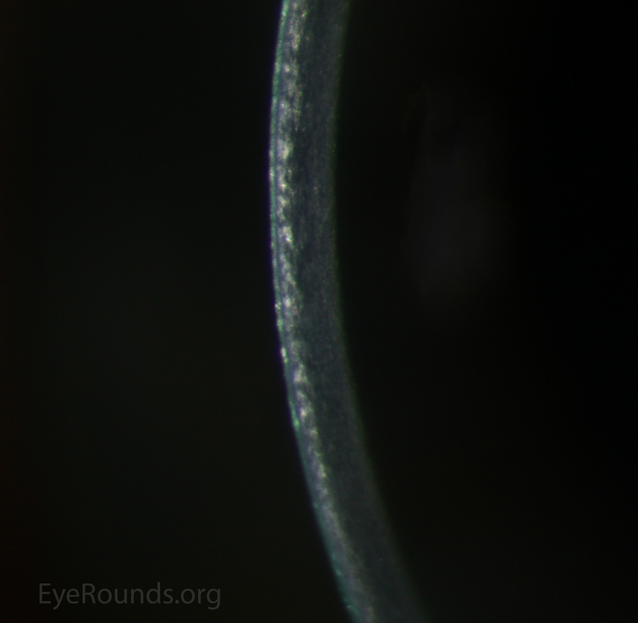
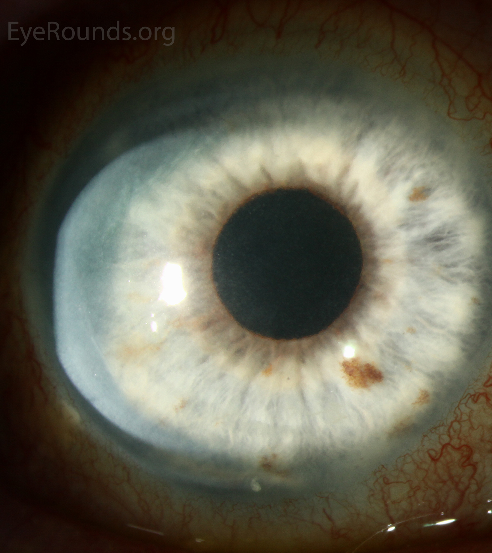
Subepithelial, or anterior stromal, corneal haze may result after superficial corneal inflammation. It can be seen as a reflective region when illuminated with a broad slit beam, but is most easily visible when using the slit beam. These photographs show the appearance of a patient with anterior stromal haze after herpes zoster virus (HZV) keratitis. The depth of the opacification is visible in the slit beam.

Corneal haze can be seen as a reflective region when illuminated with a broad slit beam
Corneal haze can be seen as a reflective region when illuminated with a broad slit beam.
Corneal haze is most easily visible when using the slit beam seen using the slit beam
Corneal haze is most easily visible when using the slit beam seen using the slit beam - Zoomed