Contributor: Ryan J. Diel, MD; Zachary Mortensen, MD; A. Tim Johnson, MD
Photographer: Antoinette Venckus, CRA
Posted January 30, 2020
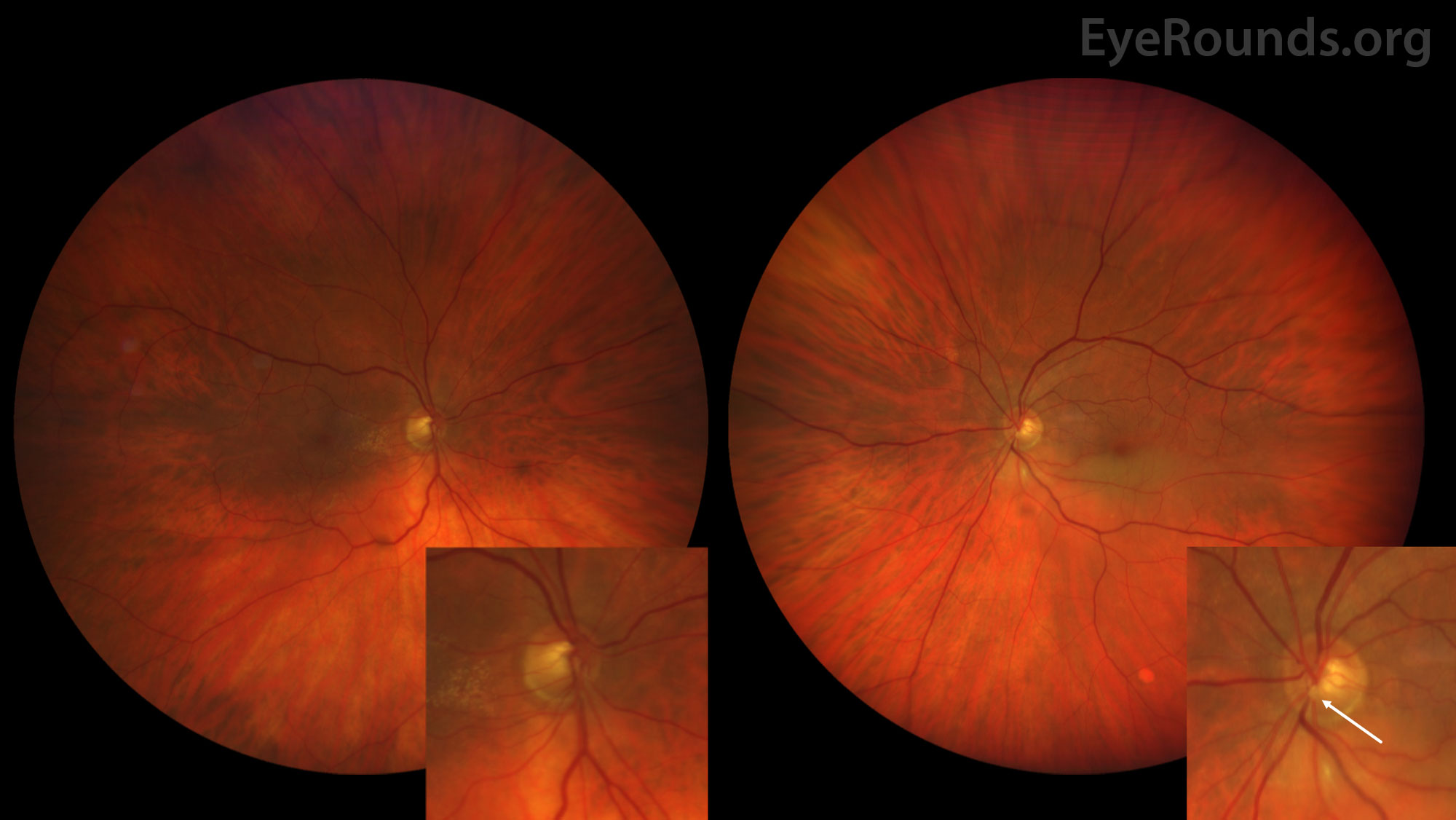
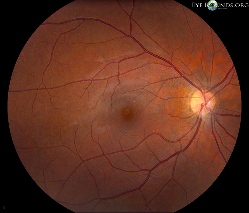
Color fundus photograph of both eyes of a 79 year-old male presenting with acute onset left eye vision loss of the superior-nasal quadrant. The right eye (left side image) demonstrates an epiretinal membrane nasal to the fovea. The left eye (right side image) demonstrates a yellow plaque (indicated by the white arrow) lodged within the inferior retinal artery overlying the optic disc. Diffuse areas of retinal whitening can be seen distal to this occlusion tracking along the inferior arcade. Optic nerve heads are magnified in the lower right corner of each image.
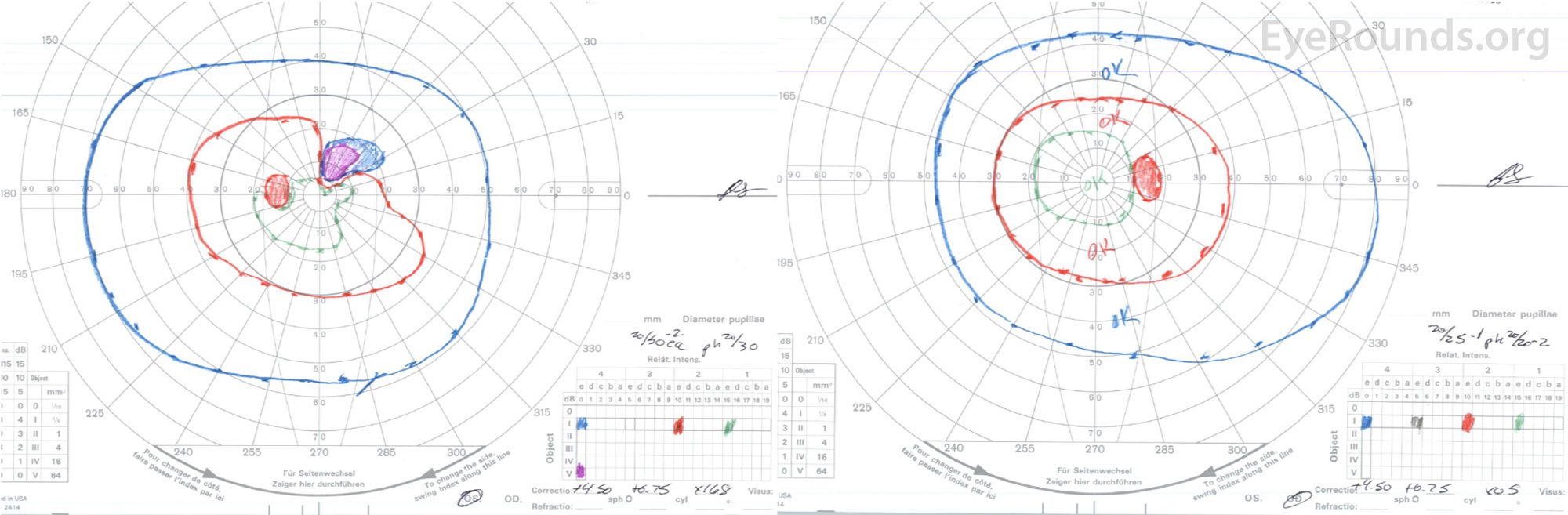
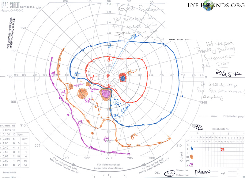
Figure 2: Goldmann visual field of both eyes. The left eye visual field (left side image) demonstrates a superiornasal scotoma which corresponds to the above mentioned Hollenhorst plaque. The right eye visual field is grossly normal.
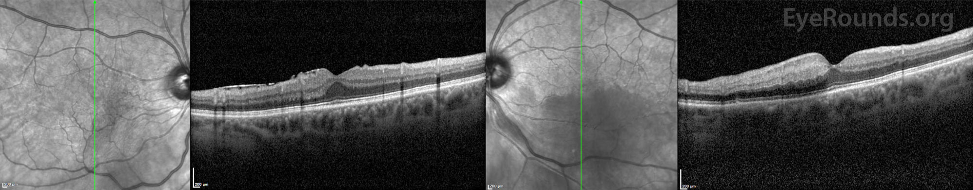
Figure 3: Optical coherence tomography (OCT) of both eyes. The right eye (left side image) is notable for the presence of an epiretinal membrane. The left eye (right side image) is significant for diffuse inner retina edema and thickening along the inferior arcade.
Contributors: John J. Chen, MD, PhD; Andrew Doan, MD, PhD; Armand P. Fasano, MD, New Jersey, Private Practice
Photographer: Cindy Montague, CRA
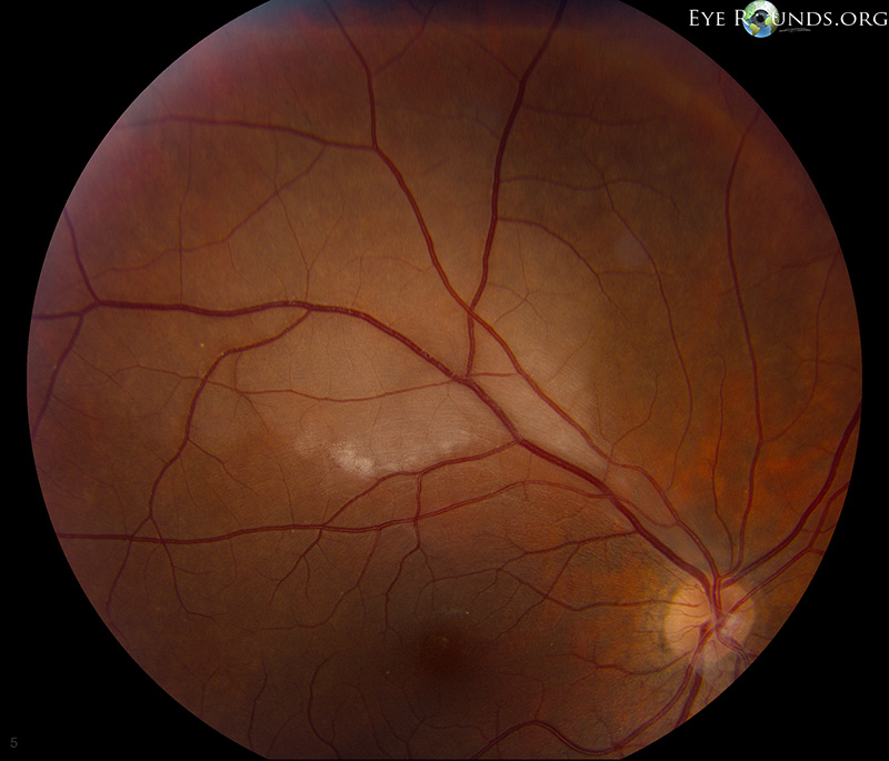
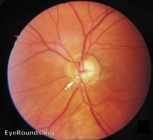
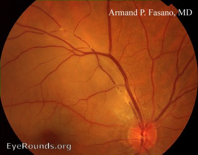
Figure 1: 31-year-old female presented with an acute onset inferonasal visual field defect in the right eye and found to have a superior branch retinal artery occlusion (Figure 1).
No emboli or Hollenhorst plaques were seen in either eye. Workup included an EKG, carotid doppler, cardiac echo, ANA, ACE, RF, CBC, PT/PTT, anticardiolipin ab, RPR, ANCA, ESR/CRP, which were all negative. (Chen, 2012)
University of Iowa
Roy J. and Lucille A. Carver College of Medicine
Department of Ophthalmology and Visual Sciences
200 Hawkins Drive
Iowa City, IA 52242
University of Iowa
Roy J. and Lucille A. Carver College of Medicine
Department of Ophthalmology and Visual Sciences
200 Hawkins Drive
Iowa City, IA 52242