Bilateral orbital disease: lymphomata
Contributor: William Charles Caccamise, Sr, MD, Retired Clinical Assistant Professor of Ophthalmology, University of Rochester School of Medicine and Dentistry
*Dr. Caccamise has very generously shared his images of patients taken while operating during the "eye season" in rural India as well as those from his private practice during the 1960's and 1970's. Many of his images are significant for their historical perspective and for techniques and conditions seen in settings in undeveloped areas.
Category: Oculoplastics

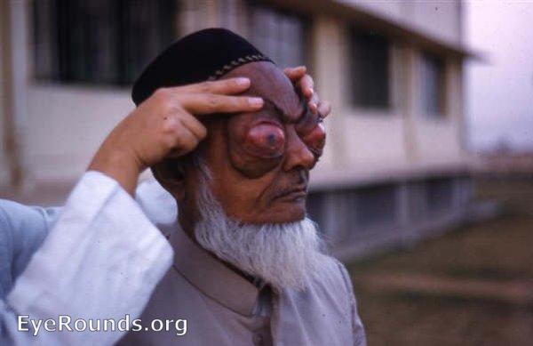
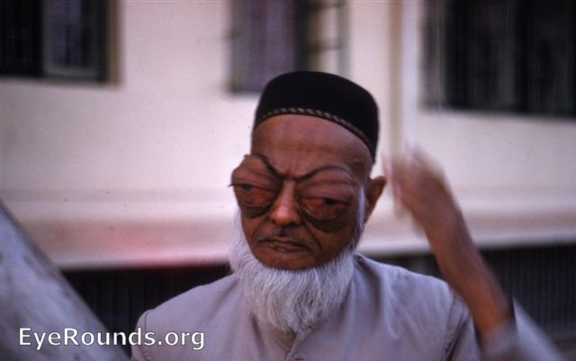
This sixtyish Indian male presented at the Kurji Holy Family Hospital Eye Clinic. The extreme condition had developed slowly over the past few years. There was still enough vision to navigate but with extreme difficulty. The patient's general health was good. The patient refused any diagnostic studies other than blood and urine analses. He refused a biopsy for orbital tissue, since he could not be assured that it would be of any practical value to him.
The clinical diagnosis was orbital lymphomata with no systemic involvement.

Frontal View of same patient

Ophthalmic Atlas Images by EyeRounds.org, The University of Iowa are licensed under a Creative Commons Attribution-NonCommercial-NoDerivs 3.0 Unported License.


