EyeRounds Online Atlas of Ophthalmology
Contributor: William Charles Caccamise, Sr, MD, Retired Clinical Assistant Professor of Ophthalmology, University of Rochester School of Medicine and Dentistry
*Dr. Caccamise has very generously shared his images of patients taken while operating during the "eye season" in rural India as well as those from his private practice during the 1960's and 1970's. Many of his images are significant for their historical perspective and for techniques and conditions seen in settings in undeveloped areas.
Category: Cataract
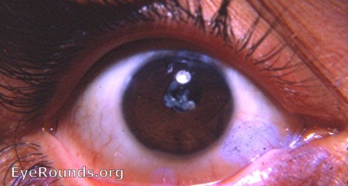
Post-needling Elschnig's pearls

As a child, this Caucasian adult had a discission (needling) of a congenital cataract. Slit-lamp examination revealed Elschnig's pearls in the pupillary area - a fish-egg-like conglomeration. The photo was taken in 1967. Reference: Yanoff and Fine, Ocular Pathology, p. 129, 1975: " Elschnig's pearls result from aberrant attempts by lens cells attached to the capsule to form new lens ' fibers '. Histologically, large clear lens cells (' bladder cells ') are seen behind the iris, in the pupillary space or in both areas."

Ophthalmic Atlas Images by EyeRounds.org, The University of Iowa are licensed under a Creative Commons Attribution-NonCommercial-NoDerivs 3.0 Unported License.


