

EyeRounds Online Atlas of Ophthalmology
Contributor: William Charles Caccamise, Sr, MD, Retired Clinical Professor of Ophthalmology, University of Rochester School of Medicine and Dentistry
*Dr. Caccamise has very generously shared his images of patients taken while operating during the "eye season" in rural India as well as those from his private practice during the 1960's and 1970's. Many of his images are significant for their historical perspective and for techniques and conditions seen in settings in undeveloped areas.
Category: External Disease
Caucasian with nevus of Ota
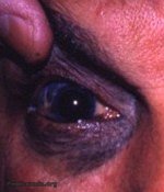
The previous photograph was of the nevus of Ota at its utmost -in an Indian. However, this photograph shows one aspect more vividly, viz. the hyperpigmentation of the skin. In lighter skinned patients as in the photograph, the increased pigmentation of the lids on the side of the lesion will be more evident than in the more heavily pigmented Indian patient. Both patients in the photographs have dark brown contralateral irides. Therefore, increased pigmentation of the involved iris can be determined only under careful evaluation - even then, it may be difficult to discern the heterochromia. It is maintained arguably by some, e.g. Yanoff and Fine in their textbook " Ocular Pathology " , that: " It is potentially malignant (uveal and skin malignant melanoma very rarely conjunctival melanoma) when it occurs in whites.
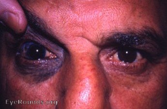
a photograph of both eyes - nevus of Ota OD

Ophthalmic Atlas Images by EyeRounds.org, The University of Iowa are licensed under a Creative Commons Attribution-NonCommercial-NoDerivs 3.0 Unported License.