This site uses tracking information. Visit our privacy policy. Click to agree to this policy and not see this again.

Ophthalmology and Visual Sciences

Confocal microscopy in fungal keratitis

Confocal microscopy in fungal keratitis

Category(ies): Cornea
Contributors: Kurt Chamberlain MS3, Justin Risma MD

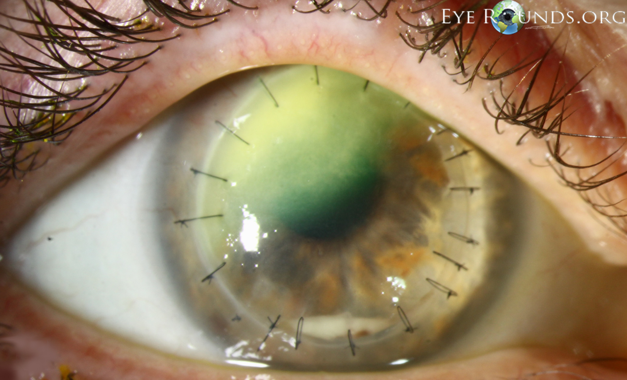
Fungal keratitis is much less common than bacterial keratitis in the United States. Risk factors for developing fungal keratitis include trauma with vegetable matter, immunosuppression (especially with topical corticosteroids), and contact lens wear. The classic appearance of fungal keratitis is a gray-white, dry-appearing infiltrate with feathery borders. However, these findings are not always present, nor are they pathognomonic for the disease, so corneal cultures, smears, and scrapings are often utilized to aid in the diagnosis.

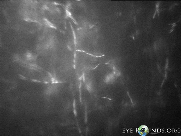
In addition to these traditional detection methods, in vivo confocal microscopy has proven to be an advantageous tool in the timely diagnosis of fungal keratitis. By providing the ability to directly visualize the presence of fungal-like structures throughout the entire depth of the cornea, confocal microscopy allows for a more rapid detection of fungi. Additionally, given the noninvasive nature of confocal microscopy, it can be used repeatedly over time to monitor response to treatment based upon the density of fungi.[1]

fungal keratitis secondary to contact lens wear
Image 1: This patient had a history of fungal keratitis secondary to contact lens wear. She was treated successfully with topical anti-fungal therapy, but developed a dense corneal scar. She underwent penetrating keratoplasty, but a few months after surgery she presented with a recurrent corneal infiltrate and a hypopyon. The diagnosis of recurrent fungal keratitis was made in part with the use of in vivo confocal microscopy.
confocal microscopy of fungal structures in the eye
Image 2: In vivo confocal microscopy image of the right cornea revealing hyper-reflective, branched, septated, linear structures suggestive of fungal structures. Fungal culture confirmed infection with fusarium species.
References:
  1. Takezawa, Y, Shiraishi A, Noda, E et al. Effectiveness of In Vivo Confocal Microscopy in Detecting Filamentous Fungi During Clinical Course of Fungal Keratitis. Cornea 2010; 29 (12): 1346-1352.
  2. Graff JM, Goins KM, Sutphin JE. Fungal Keratitis - Fusarium: 41-year-old female contact lens wearer with persisting keratitis. EyeRounds.org. date accessed: 4/3/14 ; Available from: http://www.EyeRounds.org/cases/59-fusarium-fungal-keratitis-ReNu-MoistureLoc.htm.

Image Permissions:
Creative Commons

Ophthalmic Atlas Images by EyeRounds.org, The University of Iowa are licensed under a Creative Commons Attribution-NonCommercial-NoDerivs 3.0 Unported License.