Contributor: William Charles Caccamise, Sr, MD, Retired Clinical Assistant Professor of Ophthalmology, University of Rochester School of Medicine and Dentistry
*Dr. Caccamise has very generously shared his images of patients taken while operating during the "eye season" in rural India as well as those from his private practice during the 1960's and 1970's. Many of his images are significant for their historical perspective and for techniques and conditions seen in settings in undeveloped areas.
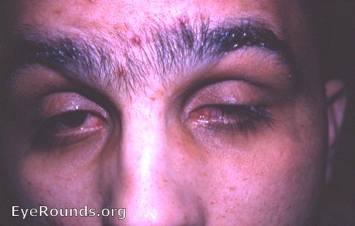
It is estimated that 25% of ptosis patients have their ptosis bilaterally. As in all cases of a congenital abnormality, ptosis cases should be evaluated for other congenital defects and a possible relationship to syndromes that include ptosis among their grouped abnormalities.
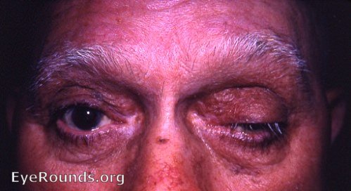
Where trachoma is endemic, a diagnosis of ptosis trachomatosa must be considered in the differential diagnosis. This patient does not have trachoma. Ptosis trachomatosa is most frequently bilateral.
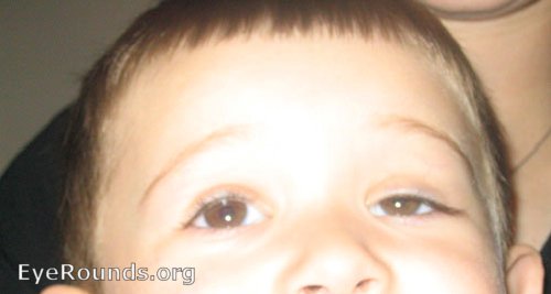
The history was that of a congenital partial ptosis OS. Examination revealed that the extraocular muscles were intact. Congenital ptosis is usually unilateral (75% of cases). It is caused by a deficiency of the striated fibers in the levator muscle.

Ophthalmic Atlas Images by EyeRounds.org, The University of Iowa are licensed under a Creative Commons Attribution-NonCommercial-NoDerivs 3.0 Unported License.


Address
University of IowaLegal
Related Links