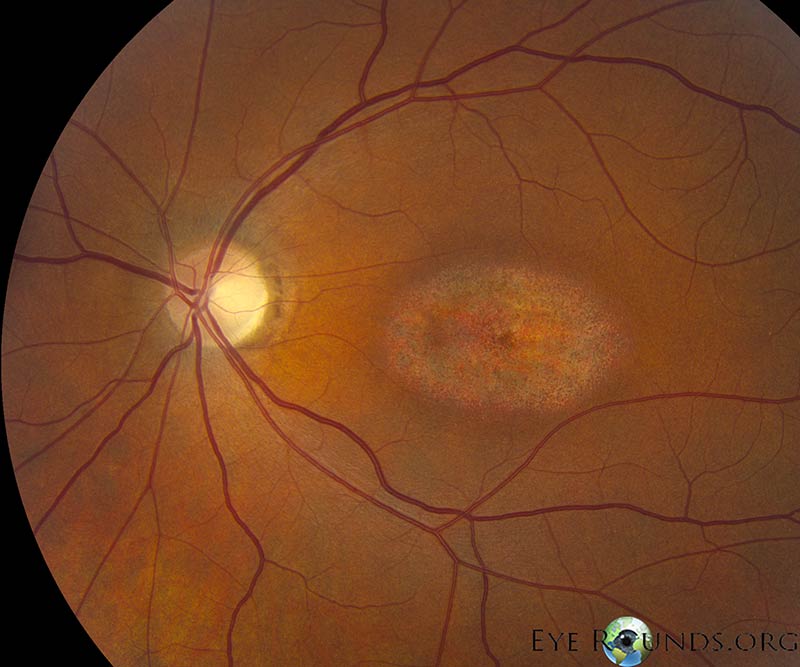
Bull's-eye maculopathy due to hydroxychloroquine toxicity
Contributor: Christopher Kirkpatrick, MD
Photographer: Stefani Karakas, CRA
June 29, 2015
Category: Retina/Vitreous
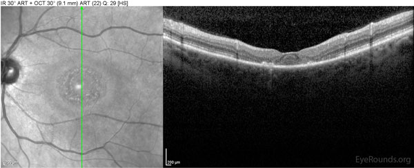
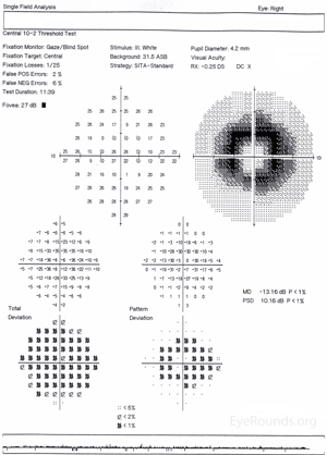
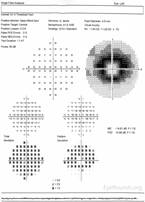
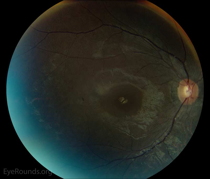
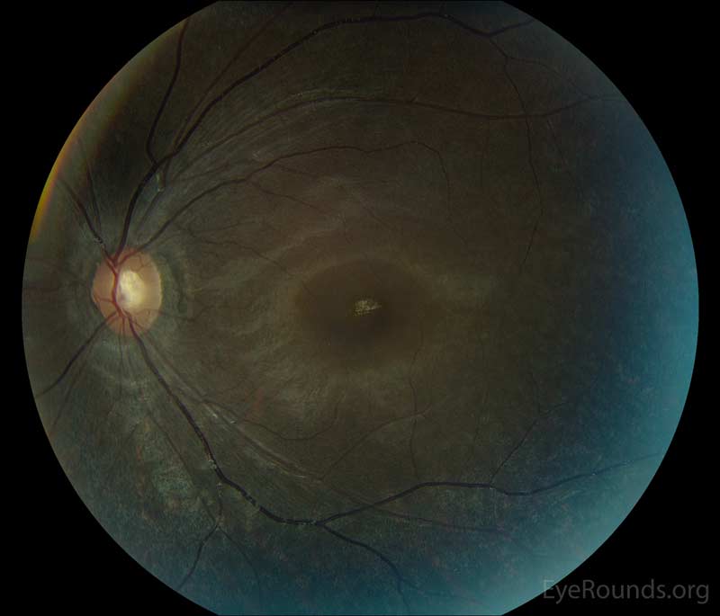
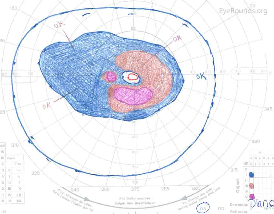
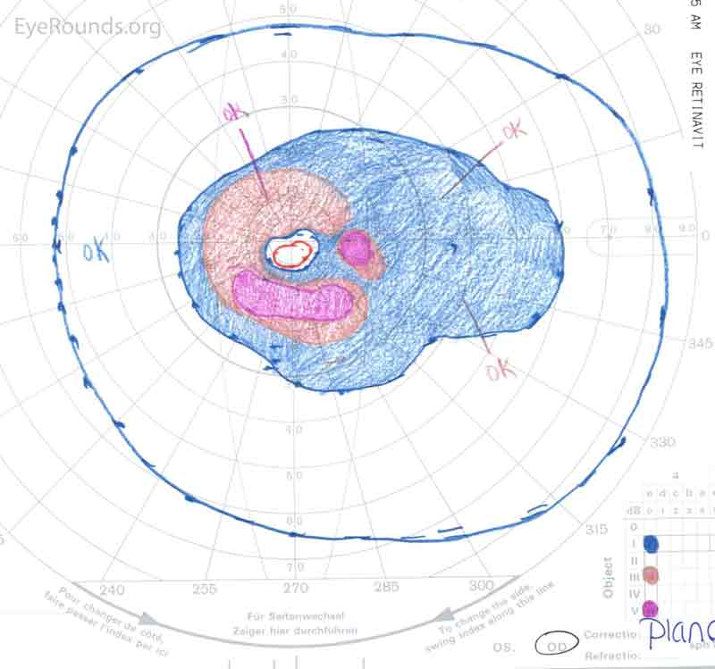
Chloroquine and hydroxychloroquine can cause toxic retinopathy due to their binding of melanin in the retinal pigmented epithelium (RPE) as well as direct toxicity to retinal ganglion cells. Early findings include mottling of the RPE and blunted foveal reflex. As the retinopathy progresses, a bull's-eye maculopathy develops, as seen in these photos. In addition to the bull's-eye maculopathy, patients can demonstrate paracentral scotomas on visual field testing and parafoveal outer retinal atrophy on OCT.
right eye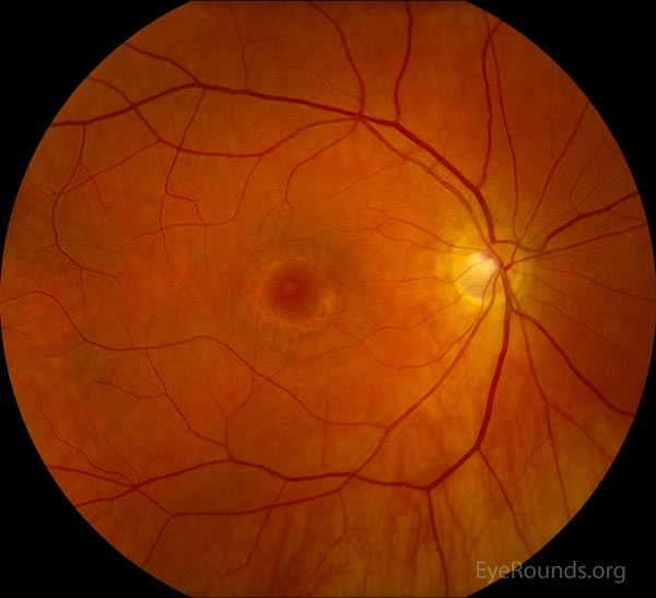 |
left eye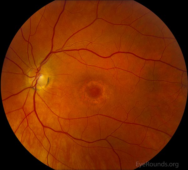 |
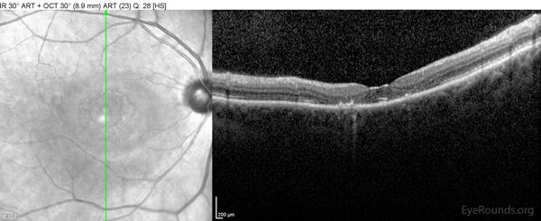 |
 |
 |
 |
Bull's-eye maculopathy due to hydroxychloroquine toxicity
Contributor: Jesse Vislisel, MD
Photographer: Randy Verdick, FOPS
September 9, 2013
This patient took hydroxychloroquine for rheumatoid arthritis and her vision dropped to 20/300 as a result of the retinal toxicity. Unfortunately, vision loss rarely recovers and the retinopathy can continue to progress even after the medication is stopped.Bull's-eye maculopathy due to hydroxychloroquine toxicity
Category(ies):
- Retina / Vitreous
Contributor: Aaron M. Ricca, MD
 |
 |
 |
 |

Ophthalmic Atlas Images by EyeRounds.org, The University of Iowa are licensed under a Creative Commons Attribution-NonCommercial-NoDerivs 3.0 Unported License.

