EyeRounds Online Atlas of Ophthalmology
Intraretinal Microvascular Abnormality (IrMA)
Contributor: Chris Kirkpatrick, MD
Photographer: Carol Chan, CRA
Category(ies): Retina/Vitreous
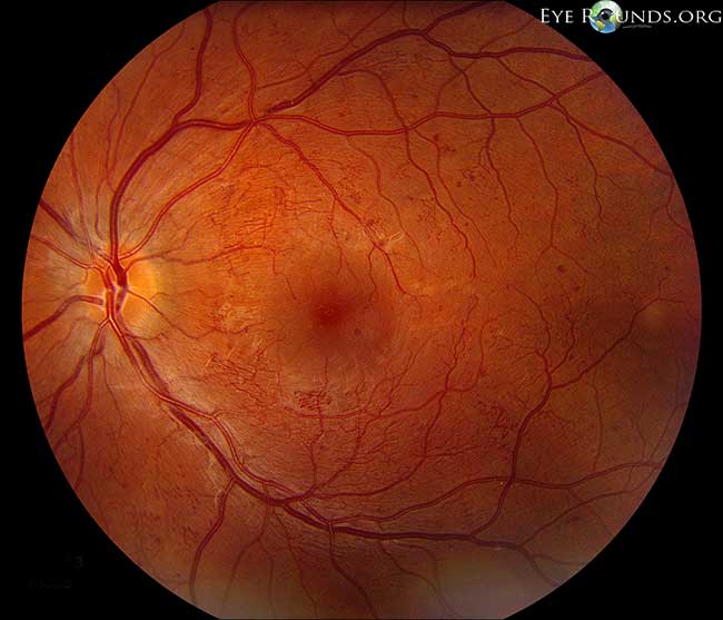
Intraretinal microvascular abnormalities (or IrMAs) are shunt vessels and appear as abnormal branching or dilation of existing blood vessels (capillaries) within the retina that act to supply areas of non-perfusion in diabetic retinopathy. These vessels represent either new vessel growth within the retina or remodeling of pre-existing vessels through endothelial cell proliferation stimulated by hypoxia bordering areas of capillary nonperfusion. IRMA is one of the defining features of severe non-proliferative diabetic retinopathy based on the "4-2-1" criteria from the Early Treatment Diabetic Retinopathy Study (ETDRS) [1]. When compared to neovascularization (NV) in proliferative disease, IRMAs are slightly larger in caliber with a more broad arrangement and are always contained to the intraretinal layers. Conversely, NV tends to be much finer and delicate in caliber, and is sometimes more focal in location depending on its severity. In severe cases, NV tends to grow along the posterior hyaloid interface especially around the optic nerve (i.e. NVD) and periphery (i.e. NVE). On fluorescein angiography, NV will often show late leakage whereas IRMAs traditionally do not leak.
This picture represents a young insulin dependent diabetic patient with severe non-proliferative diabetic retinopathy. There is great representation of IRMA perifoveally, but most impressively just inferior to the fovea.
Reference
1. Gangnon RE, Davis MD, Hubbard LD, Aiello LM, Chew EY, Ferris FL 3rd, Fisher MR; Early Treatment Diabetic Retinopathy Study Research Group. A severity scale for diabetic macular edema developed from ETDRS data. Invest Ophthalmol Vis Sci. 2008 Nov;49(11):5041-7.

Ophthalmic Atlas Images by EyeRounds.org, The University of Iowa are licensed under a Creative Commons Attribution-NonCommercial-NoDerivs 3.0 Unported License.


