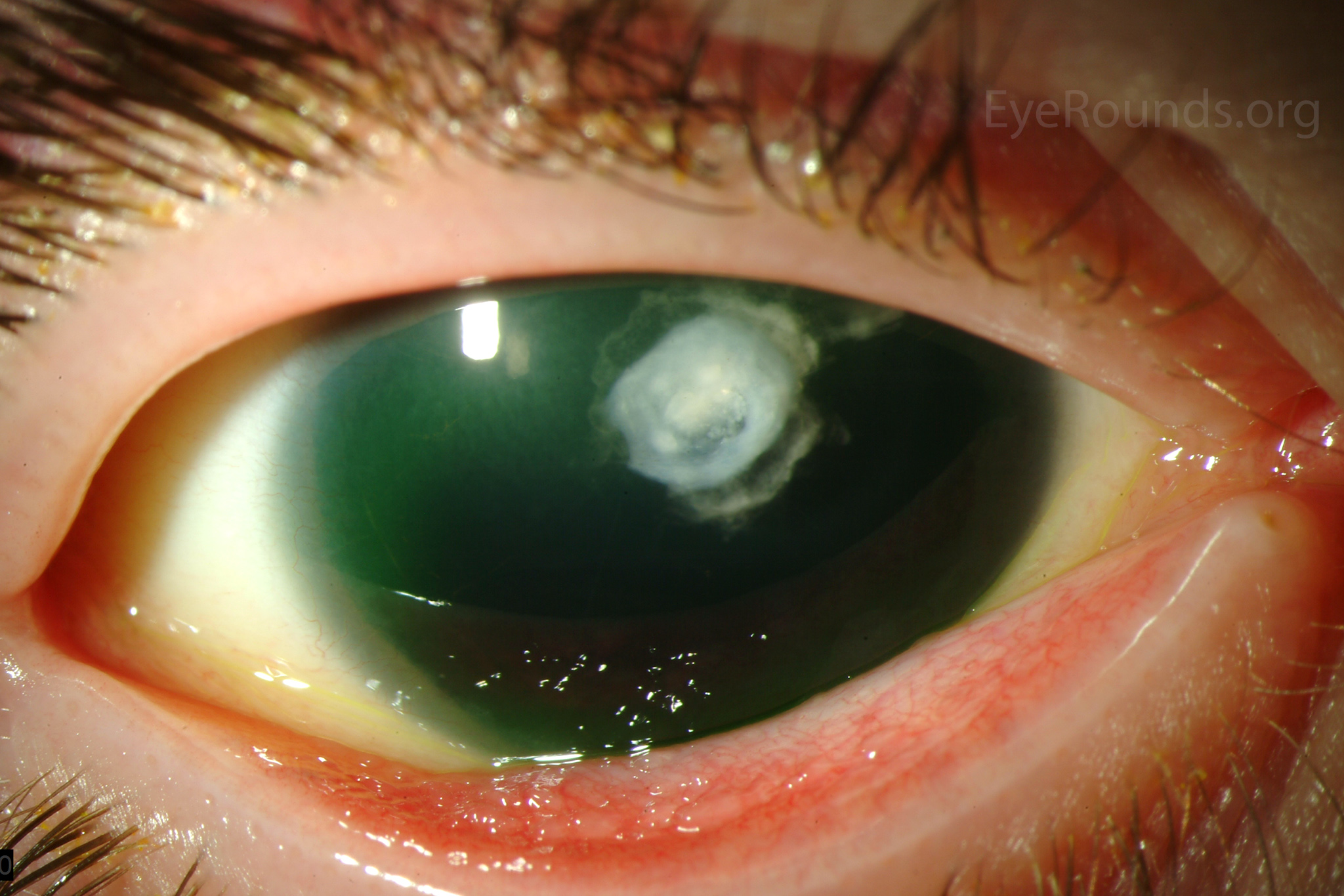
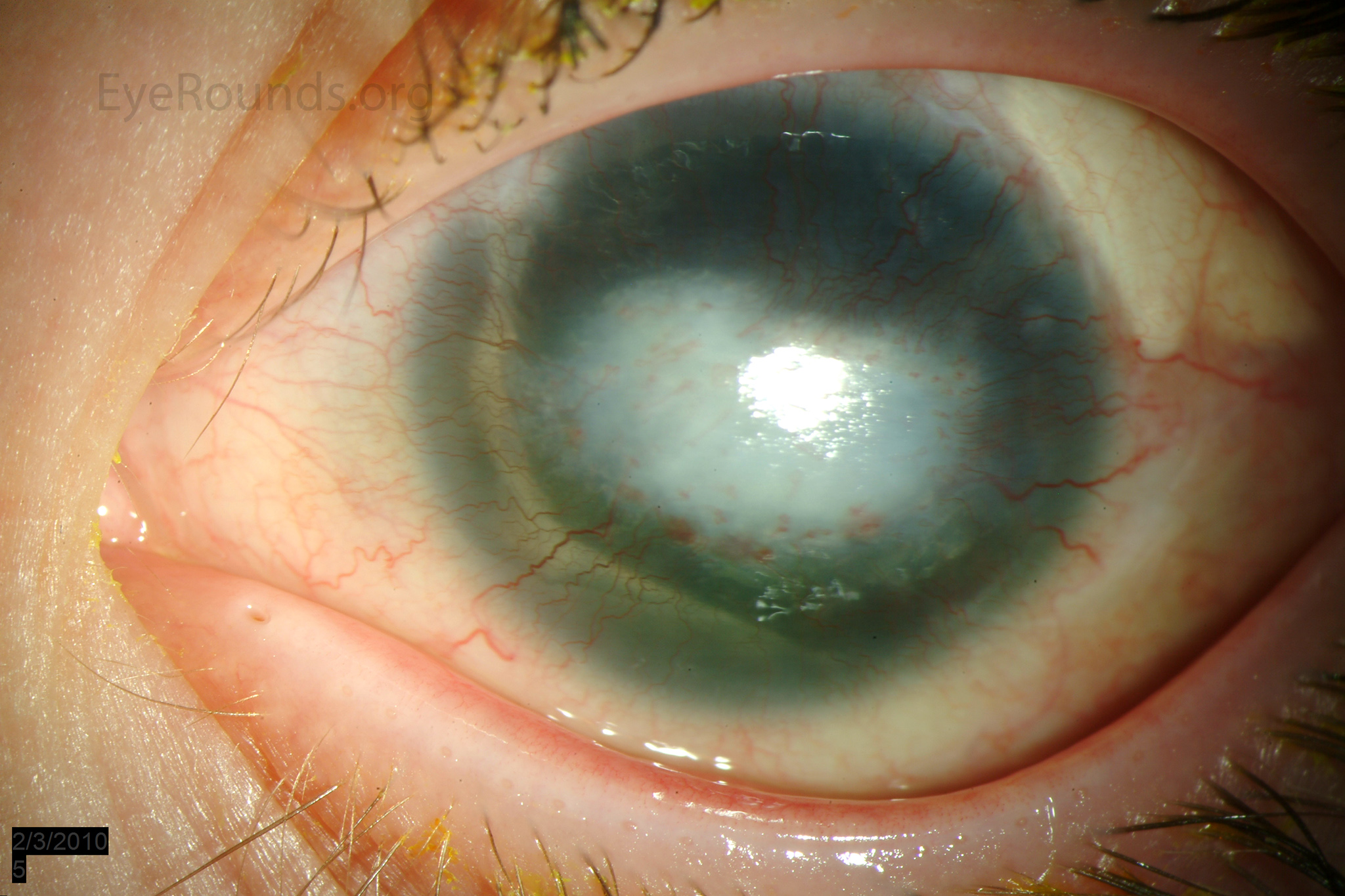
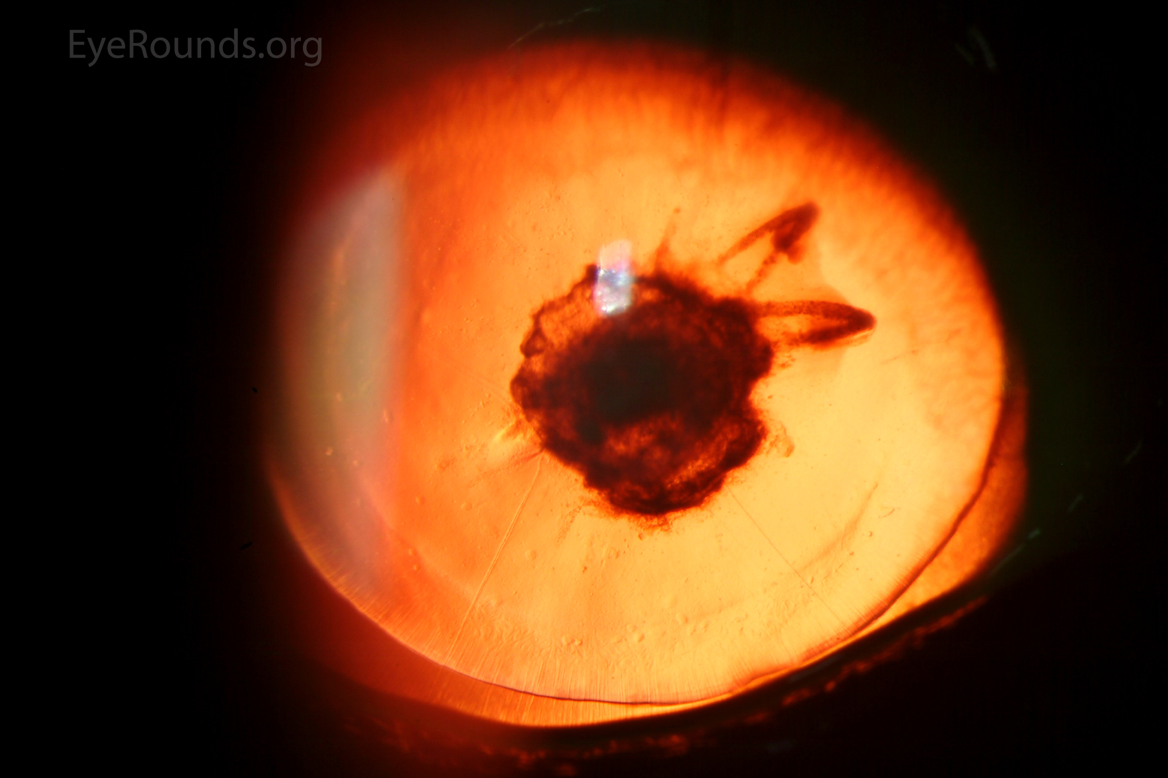
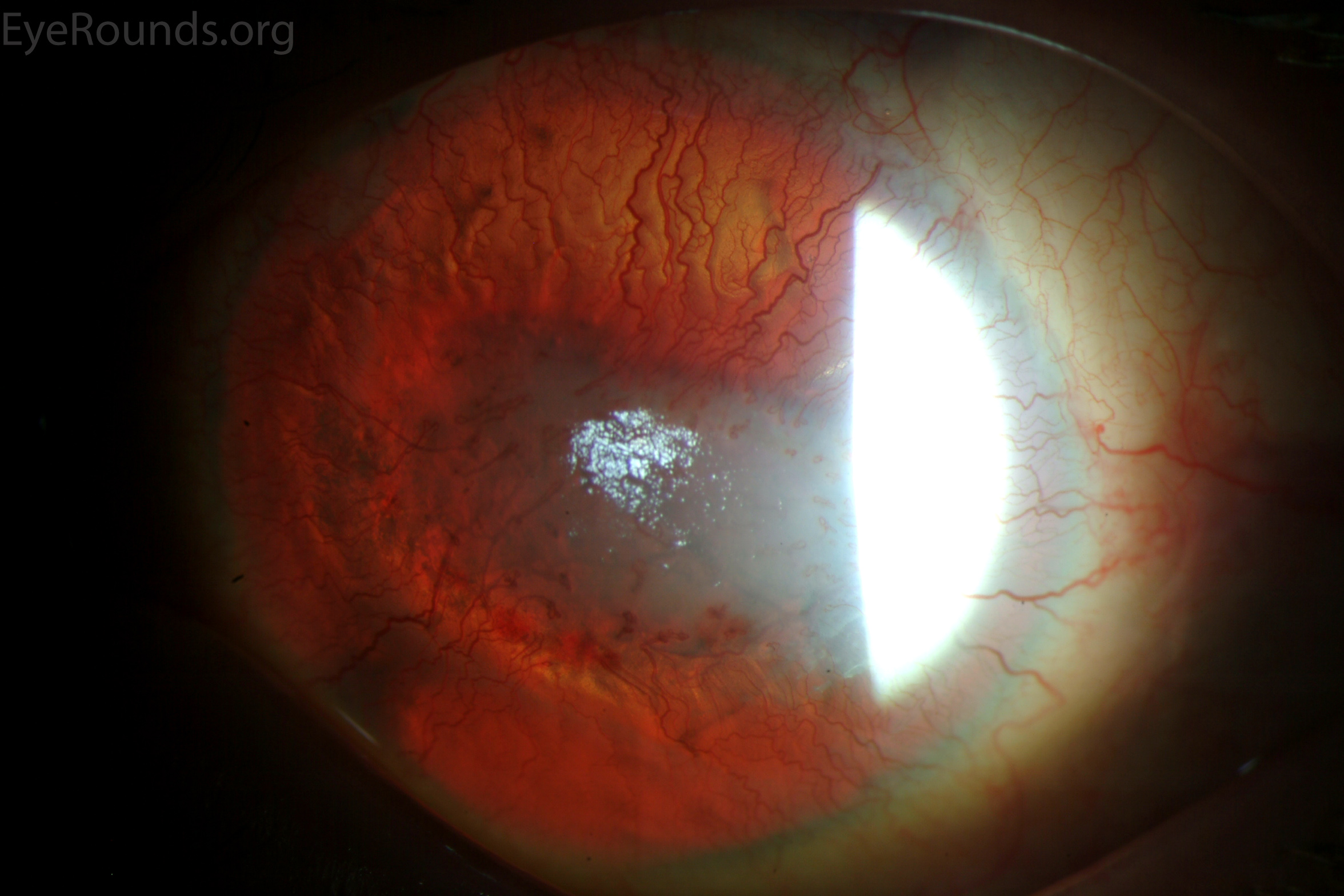
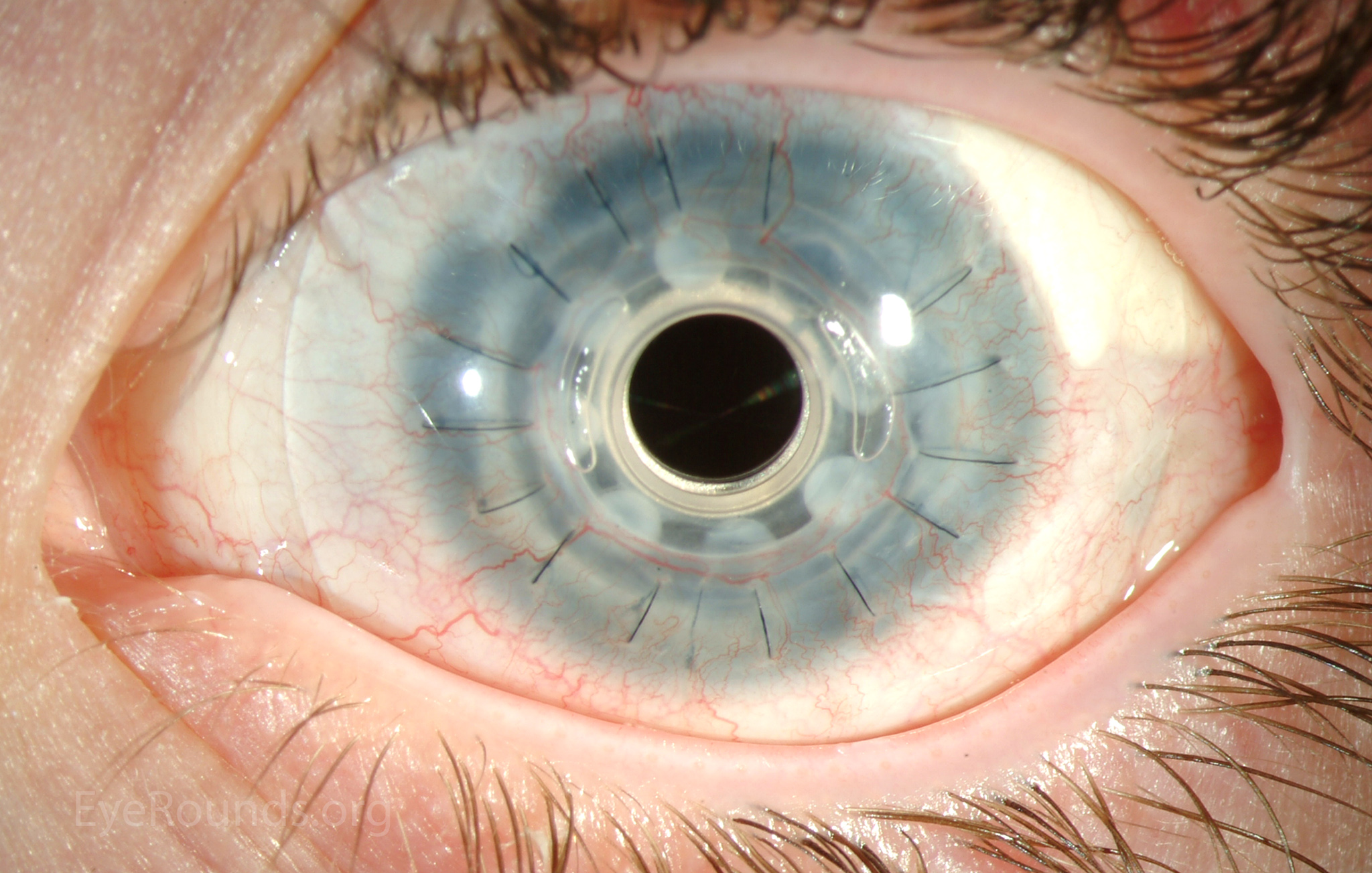
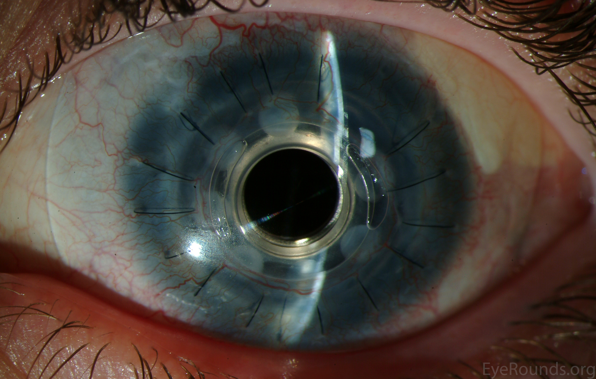
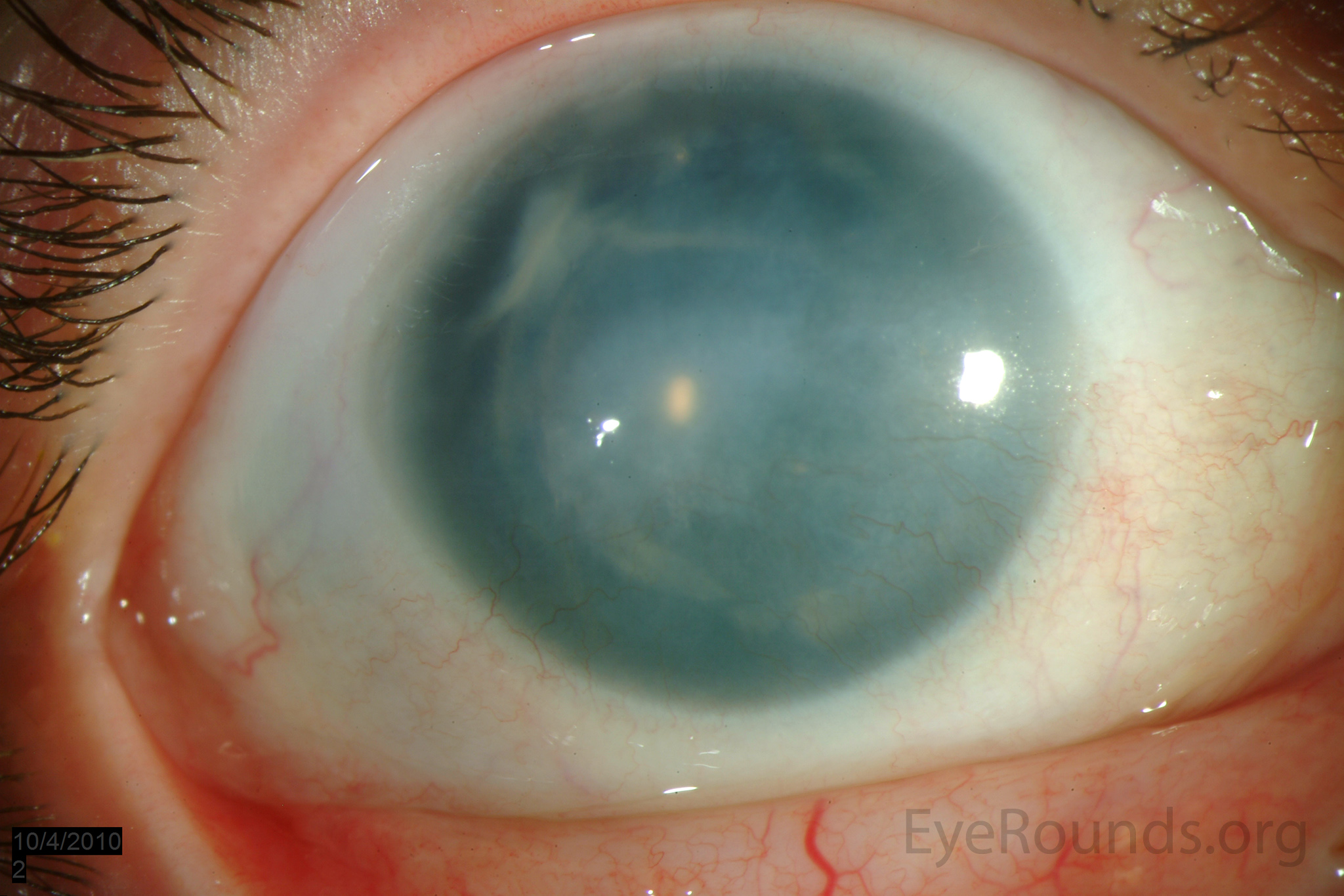
Congenital aniridia is a hereditary disease most commonly with autosomal dominant inheritance. These slit lamp photographs of a 12-year-old with congenital aniridia (Fig. 1 A, B) show conjunctivalization of the left cornea due to stem cell deficiency and a central corneal scar. There is iris hypoplasia in both eyes. The patient has a congenital cataract in the right eye and is aphakic in the left eye after having undergone cataract surgery. The cataract and corneal scars are more easily demonstrated on retroillumination. Also note the visible inferior border of the crystalline lens and zonules in the right eye, a feature not visible in individuals with normal irides. The lower photos (Fig. 2) show the appearance of the left eye after placement of a Boston Keratoprosthesis (KPro). Please see the related case report for more information about congenital aniridia.

Ophthalmic Atlas Images by EyeRounds.org, The University of Iowa are licensed under a Creative Commons Attribution-NonCommercial-NoDerivs 3.0 Unported License.

Address
University of IowaLegal
Related Links