

Lacrimal gland tumor
Contributor: William Charles Caccamise, Sr, MD, Retired Clinical Professor of Ophthalmology, University of Rochester School of Medicine and Dentistry
*Dr. Caccamise has very generously shared his images of patients taken while operating during the "eye season" in rural India as well as those from his private practice during the 1960's and 1970's. Many of his images are significant for their historical perspective and for techniques and conditions seen in settings in undeveloped areas.
This female Indian patient presented with this lacrimal gland lesion. The differential diagnosis includes benign mixed tumors, malignant epithelial tumors, and nonepithelial tumors. Approximately 50% of lacrimal gland tumors are epithelial and approximately 50% are lymphoid tumors and inflammatory psudotumors. Benign mixed tumor is the most common benign neoplasm of the lacrimal gland.
Lacrimal gland tumor with medial displacement of eyeball

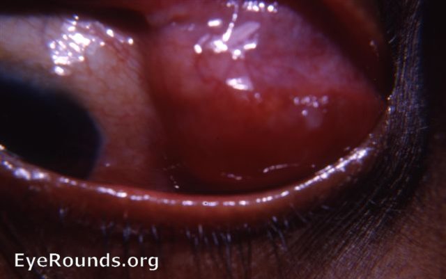
Lacrimal gland tumor, mixed type
Elevation of the left upper lid revealed a tumor of the left tear gland. Lack of tenderness and a long, benign history pointed towards mixed tumor rather than a dacryoadenitis.Biopsies are contraindicated in cases of suspected mixed tumors.

Elevation of the lid reveals a large lacrimal gland tumor.

Elevation of the upper lid reveals a large lacrimal gland tumor - mixed type

Ophthalmic Atlas Images by EyeRounds.org, The University of Iowa are licensed under a Creative Commons Attribution-NonCommercial-NoDerivs 3.0 Unported License.