This site uses tracking information. Visit our privacy policy. Click to agree to this policy and not see this again.

Ophthalmology and Visual Sciences

Multiple evanescent white dot syndrome (MEWDS)

Multiple evanescent white dot syndrome (MEWDS)

Category(ies): Retina, Uveitis
Contributor: Jesse Vislisel, MD; Karen Gehrs, MD
Photographer: Carol Chan, CRA

Figure 1

Multiple evanescent white dot syndrome (MEWDS) results from idiopathic inflammation of the retina. It is most common in healthy, young, white, myopic females and typically has an acute and unilateral presentation.

Multiple evanescent white dot syndrome (MEWDS) fundus
As seen in the photograph, the condition causes multiple, discrete, gray-white spots in the outer retina or retinal pigmented epithelium (RPE). There are pathognomonic granular changes within the fovea. Mild intraocular inflammation may be present.
Multiple evanescent white dot syndrome (MEWDS) Fundus fluorescein angiography (FFA)
Fundus fluorescein angiography (FFA) shows early punctate hyperfluorescence
Multiple evanescent white dot syndrome (MEWDS) Fundus fluorescein angiography (FFA) late
Fundus fluorescein angiography (FFA) shows late staining of the lesions arranged in a "wreath-like" configuration around the fovea.

Figure 2
Contributor: Jeffrey Welder, MD
Photographer: : Brice Critser, CRA
Multiple evanescent white dot syndrome (MEWDS) fundus
A 25-year-old male presented with 3 days of painless scotoma and photopsia in the left eye. Amsler grid testing revealed an enlarged blindspot. Fundus examination showed multiple subretintal white spots. Fluorescein angiography showed classic punctate hyperfluorescence in a wreath-like pattern in the left eye (photo at bottom of page). A diagnosis of MEWDS was made.
Multiple evanescent white dot syndrome (MEWDS) fundus normal
Fundus, right (normal)
Multiple evanescent white dot syndrome (MEWDS) fundus abnormal with white spots
Fundus, eft (abnormal)
Multiple evanescent white dot syndrome (MEWDS) fundus FA
Fluorescein angiography showed classic punctate hyperfluorescence in a wreath-like pattern in the left eye

Figure 3
Contributor: Luke Lenci, MD
Photographer: : Brice Critser, CRA

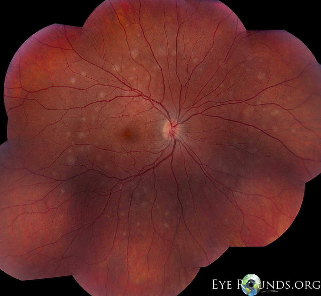
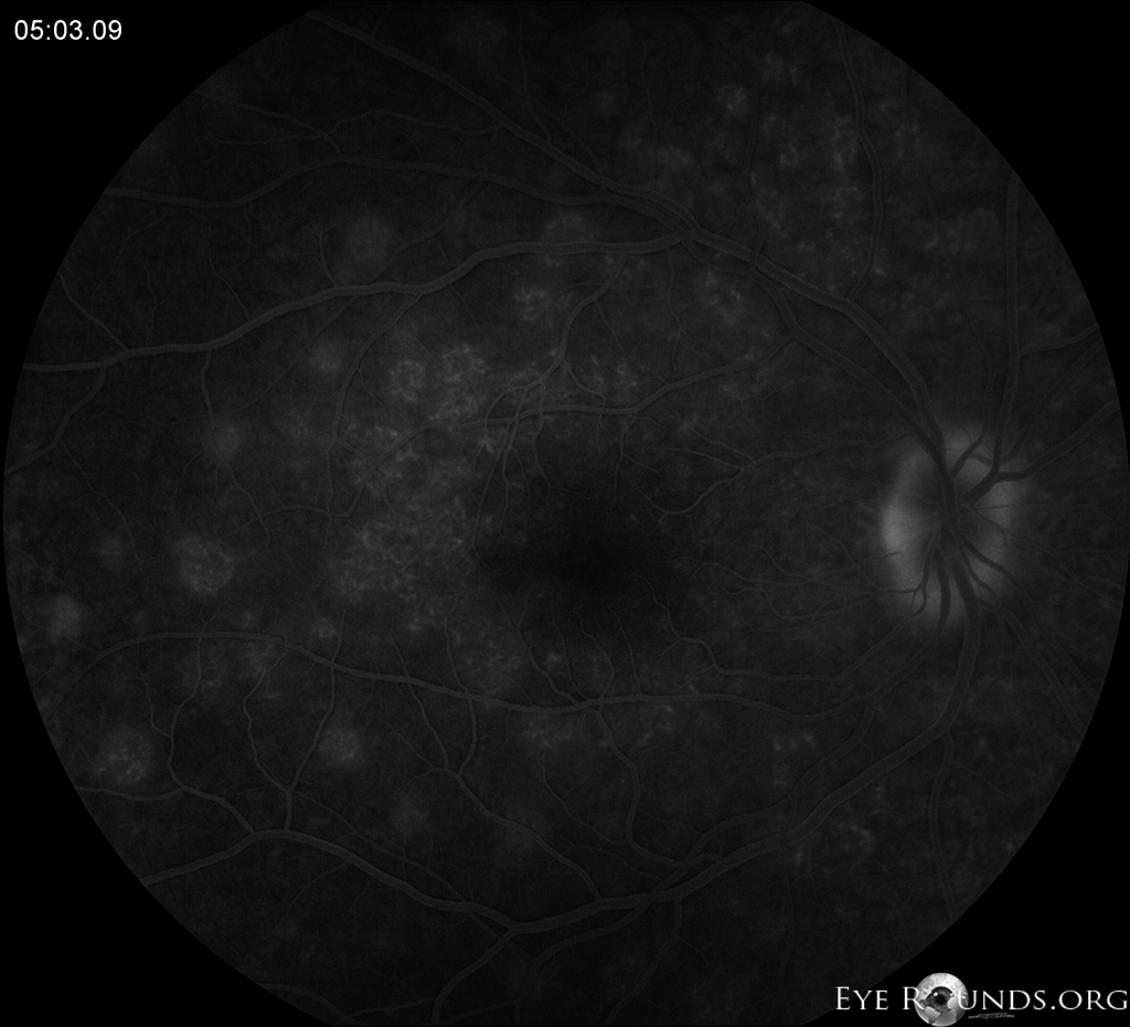
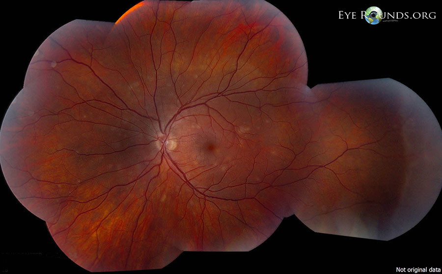
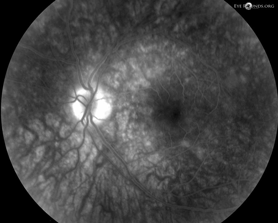
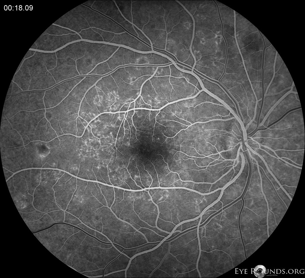
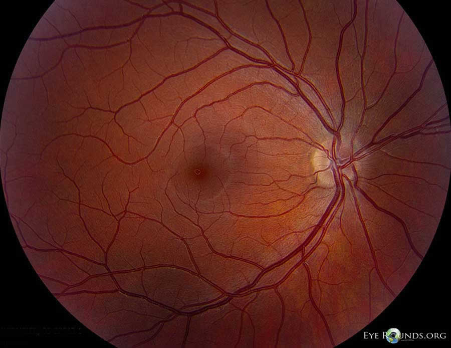
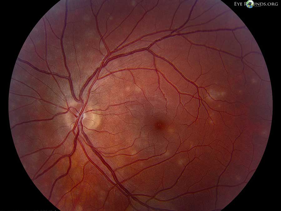
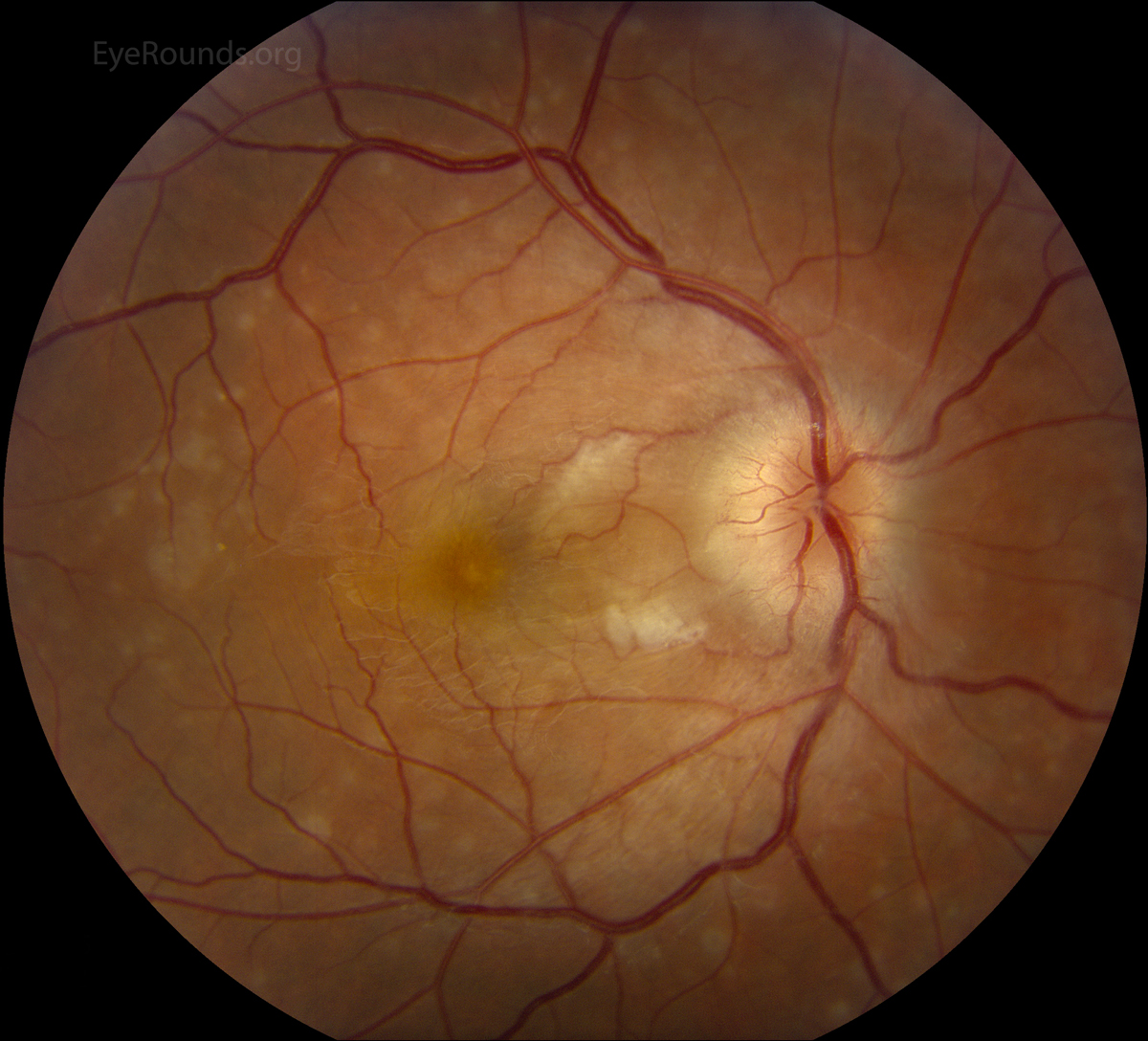
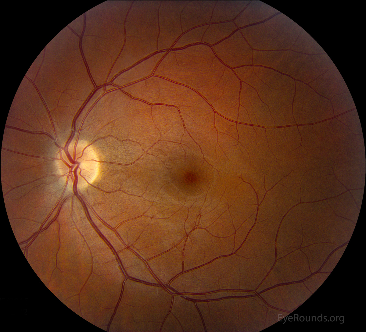
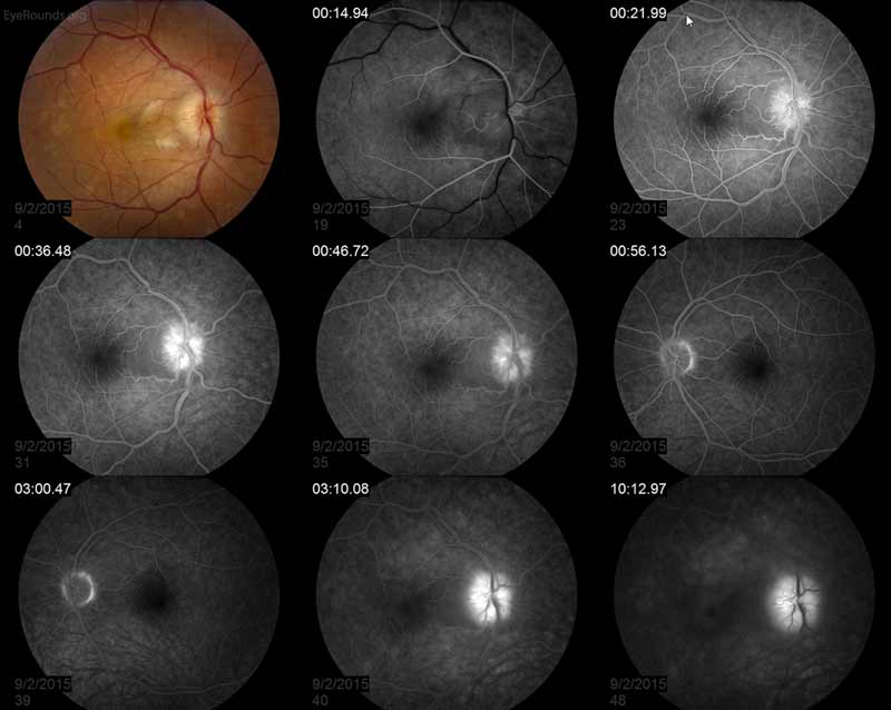
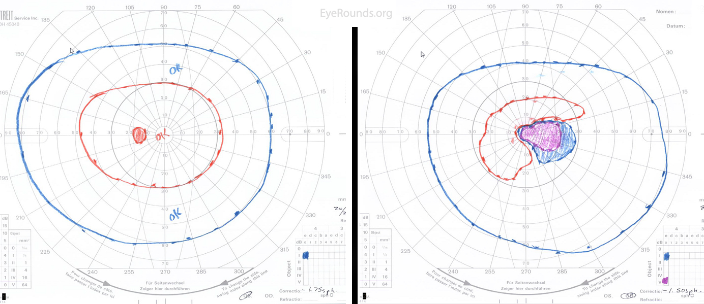
This case represents the more severe end of the spectrum for MEWDS. This young, myopic female presented with a complaint of unilateral shimmering photopsias and a paracentral scotoma. Her exam showed the pathognomic subfoveal yellow/orange granularity in the right eye along with an FFA that had a classic "wreathlike" hyperfluorescence. Disc edema is also seen in the right eye.