EyeRounds Online Atlas of Ophthalmology
Contributor: William Charles Caccamise, Sr, MD, Retired Clinical Professor of Ophthalmology, University of Rochester School of Medicine and Dentistry
*Dr. Caccamise has very generously shared his images of patients taken while operating during the "eye season" in rural India as well as those from his private practice during the 1960's and 1970's. Many of his images are significant for their historical perspective and for techniques and conditions seen in settings in undeveloped areas.
Category: Cataract
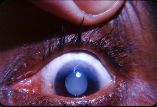
Pannus -superior limbus with Morgagnian cataract

The white pupil is due to a Morgagnian cataract. The classic floating nucleus is hidden by the white liquefied cortex. Visibility of the nucleus is strictly a positional matter. Close observation will reveal white dots in some periperal areas of the cataract. Pannus of trachoma reveals itself by the fine blood vessels invading the cornea superiorly. The definition of pannus of the cornea is: young vascularized connective tissue (granulation tissue) that is growing into the cornea. In trachoma it normally involves the superior limbus. While Dr. Caccamise was Public Health Officer of Chiba Prefecture, Japan,in 1949,and associated with the eye department of Chiba Medical College, an entire grammar school population was evaluated for trachoma. Each child in turn sat at a slit-lamp and was examined for pannus. The absence or presence of blood vessel invasion of the superior cornea was the essential finding in screening the children.


Ophthalmic Atlas Images by EyeRounds.org, The University of Iowa are licensed under a Creative Commons Attribution-NonCommercial-NoDerivs 3.0 Unported License.


