This site uses tracking information. Visit our privacy policy. Click to agree to this policy and not see this again.

Ophthalmology and Visual Sciences

High peripheral anterior synechia in iridocorneal endothelial syndrome

High peripheral anterior synechia in iridocorneal endothelial syndrome

Category(ies): Glaucoma/iris
Contributor: Justin Risma, MD
Photographer: Brice Critser, CRA

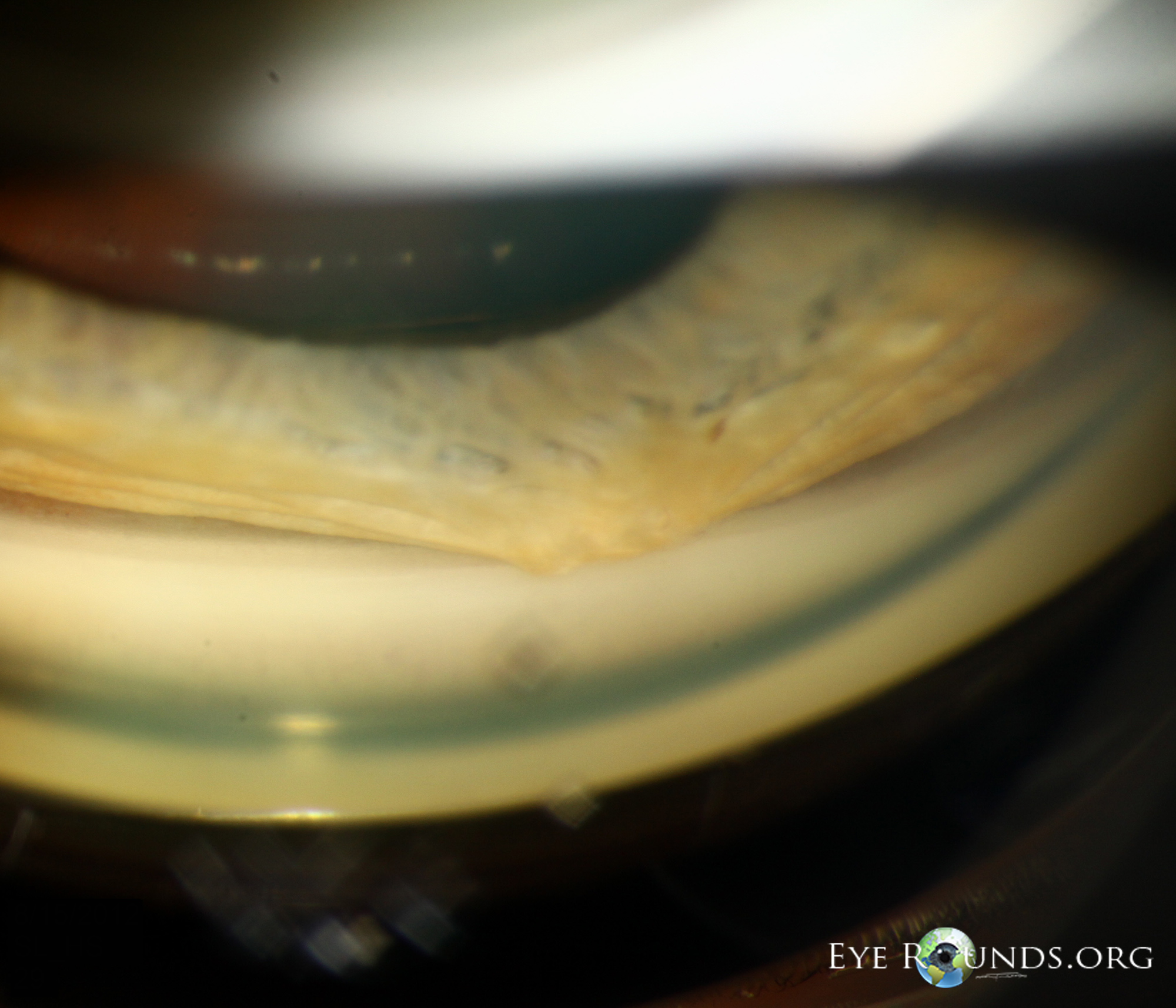
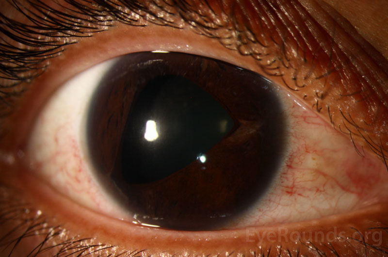
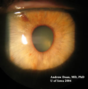
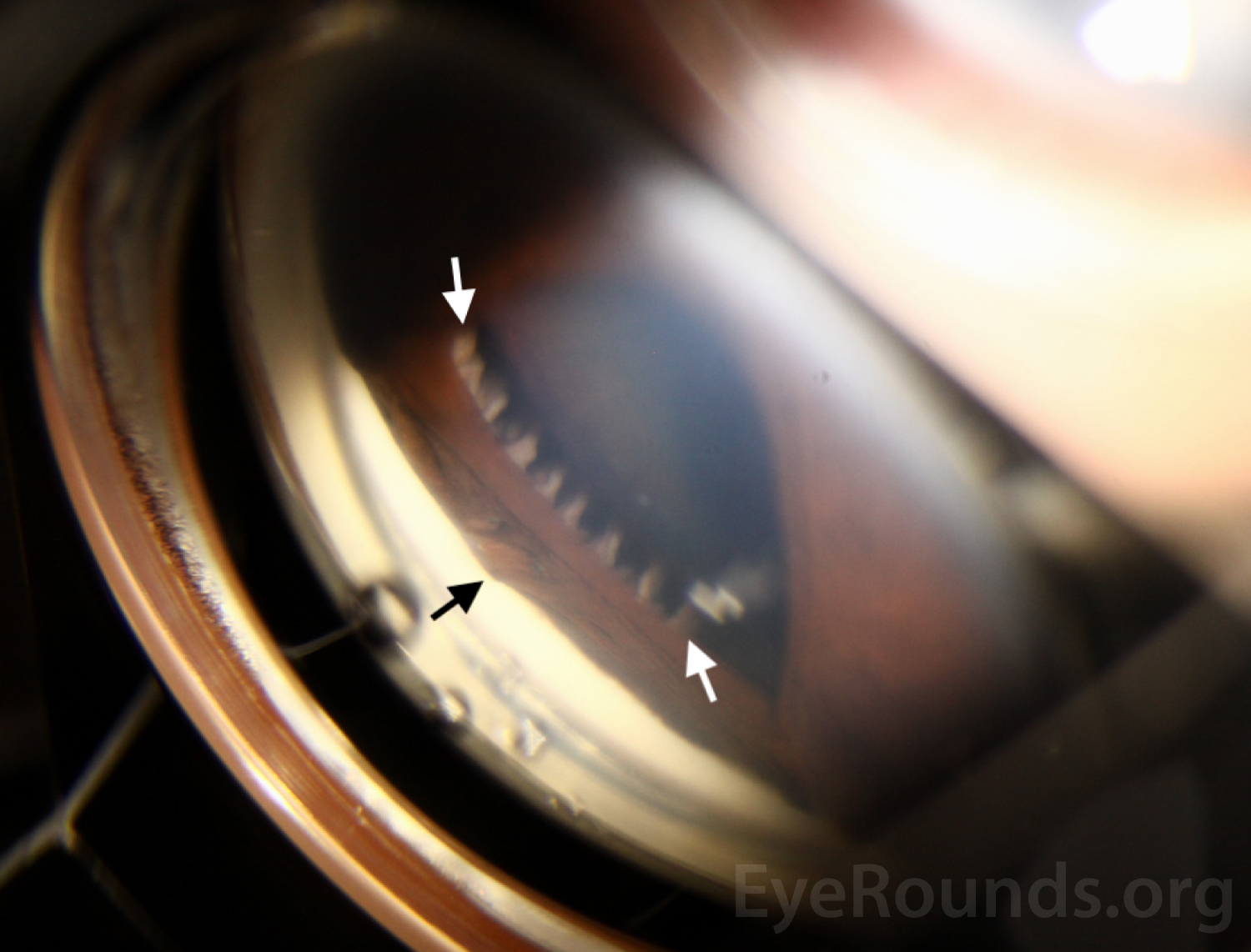
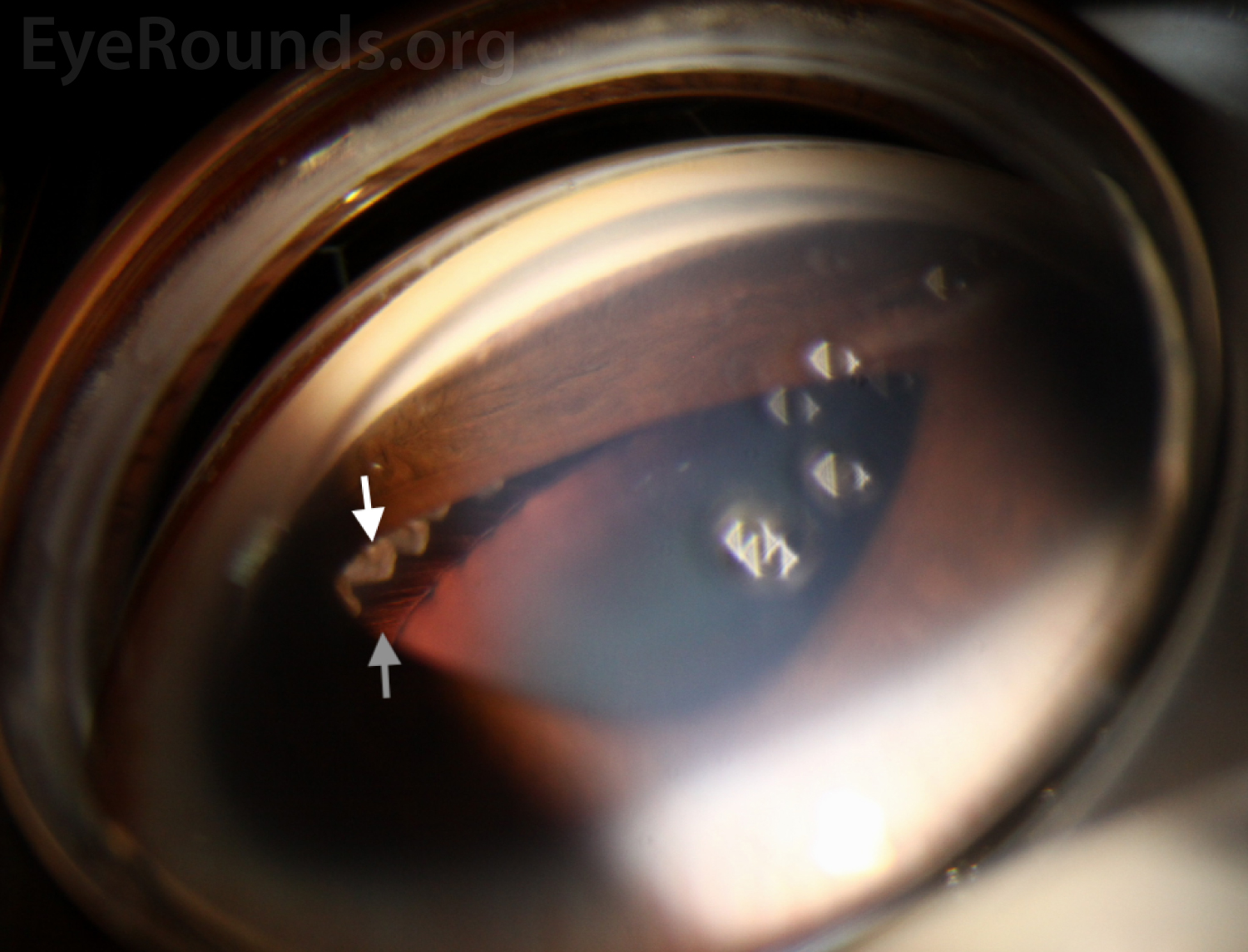
Peripheral anterior synechiae (PAS) are adhesions of the peripheral iris to the structures in the angle of the anterior chamber. They may be associated with a variety of conditions such as angle closure glaucoma, neovascular glaucoma, uveitis, and iridocorneal endothelial (ICE) syndrome.

gonioscopic photo demonstrates a high PAS which extends to Schwalbe's line and is characteristic of ICE syndrome
This gonioscopic photo demonstrates a high PAS which extends to Schwalbe's line and is characteristic of ICE syndrome.

High peripheral anterior synechia in iridocorneal endothelial syndrome

High peripheral anterior synechia in iridocorneal endothelial syndrome

Category(ies): Glaucoma/iris
Contributor: Andrew Goldstein, MD; and Erin Boese, MD
Photographer: Jody Troyer, CRA

This series of images was obtained in a 30-year-old female who presented for evaluation of an abnormal pupil and was subsequently diagnosed with iridocorneal endothelial (ICE) syndrome.

anterior segment of the right eye shows corectopia with the pupil drawn inferotemporally.
Figure 1: Color photography of the anterior segment of the right eye shows corectopia with the pupil drawn inferotemporally.
 Gonioscopic photography of the right eye shows high peripheral anterior synechiae (PAS)
Figure 2: Gonioscopic photography of the right eye shows high peripheral anterior synechiae (PAS) noted by the black arrow and visible ciliary processes (between the white arrows).
 Gonioscopic photography of the right eye provides a unique view of the ciliary processes (white arrow) and zonules of the crystalline lens
Figure 3: Gonioscopic photography of the right eye provides a unique view of the ciliary processes (white arrow) and zonules of the crystalline lens (grey arrow) due to high PAS.

Related Articles