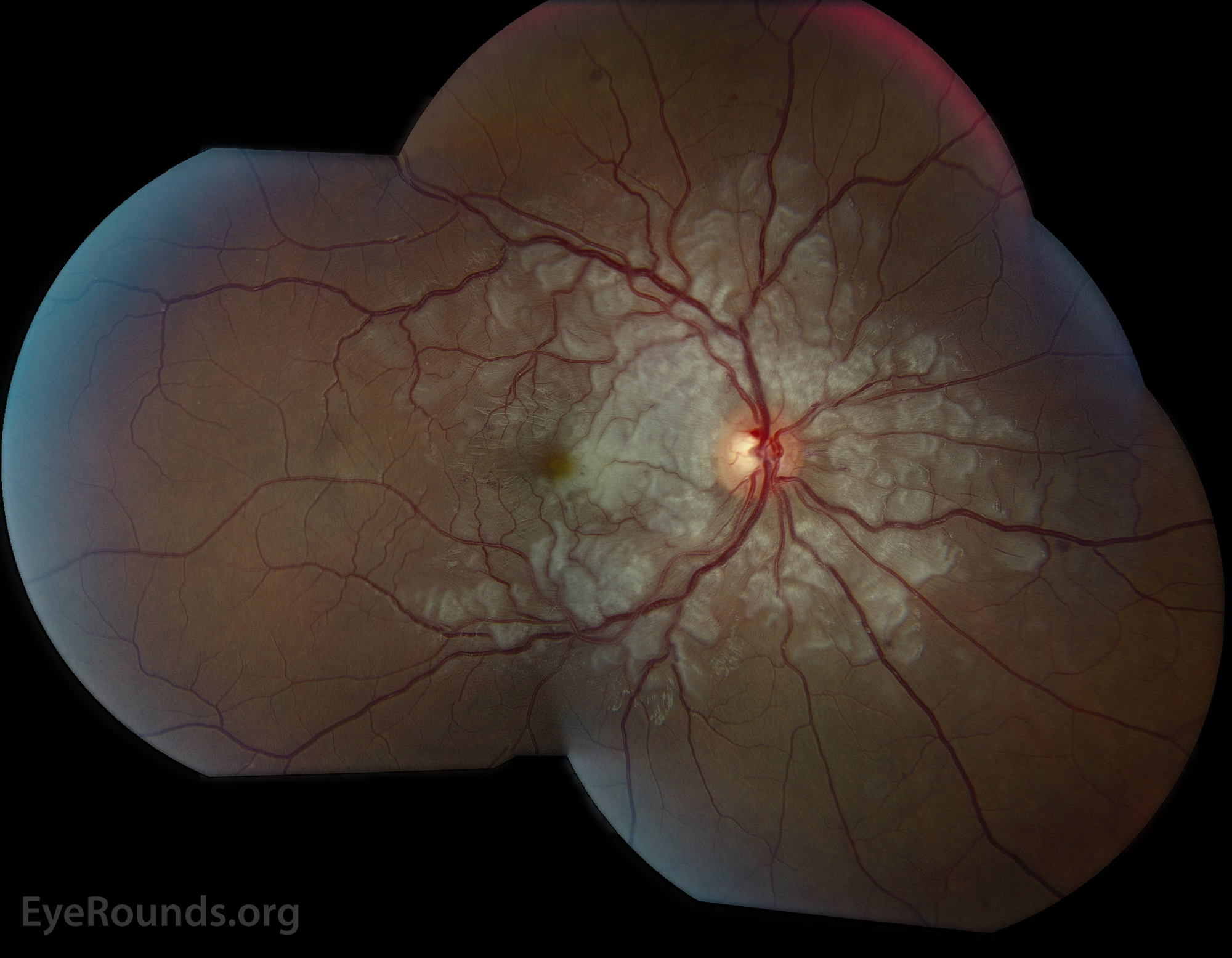
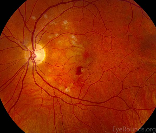
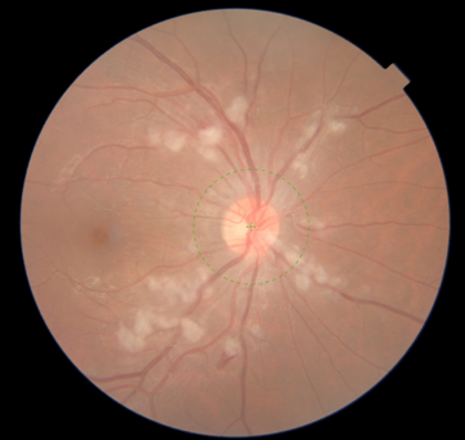
This 21 year old male noted a significant unilateral decrease in vision two days after an all-terrain vehicle (ATV) roll-over accident in which he suffered head trauma and a fractured rib. Radiating superficial retinal ischemia is seen here with scattered blot hemorrhages and venous dilation in the right eye. His left eye was unaffected. True Purtscher's Retinopathy was originally described in cases of known head-trauma, but this clinical picture can also be seen in a variety of disease mechanisms including chest compression, long-bone fracture, acute pancreatitis, and connective tissue disorders amongst others.
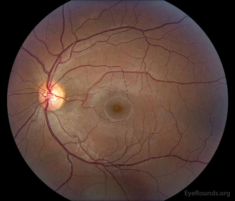
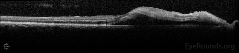
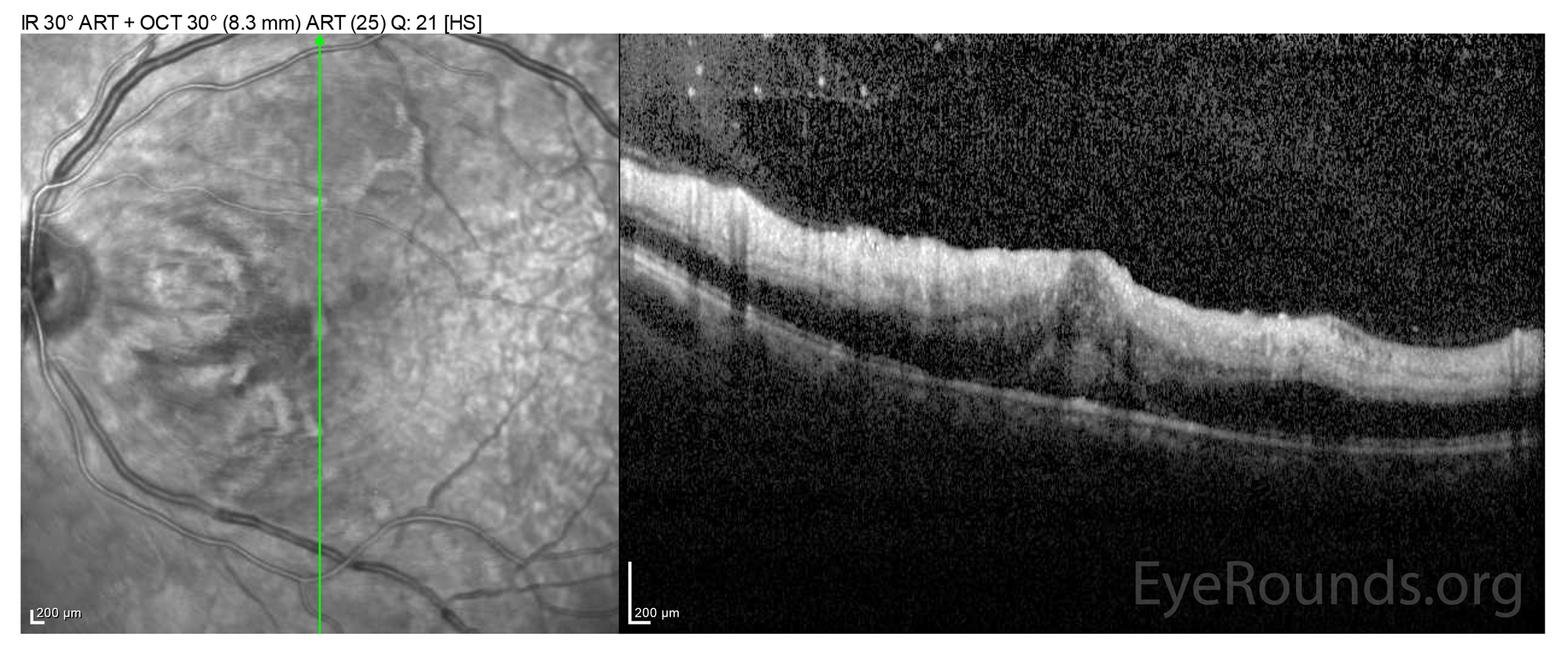
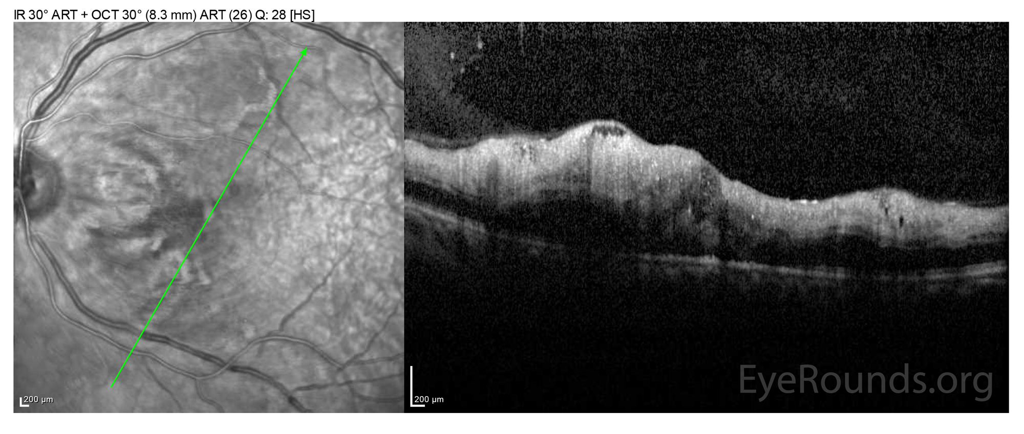
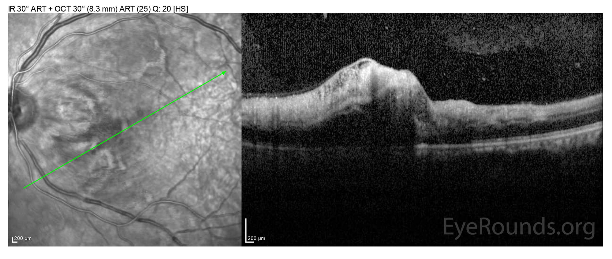
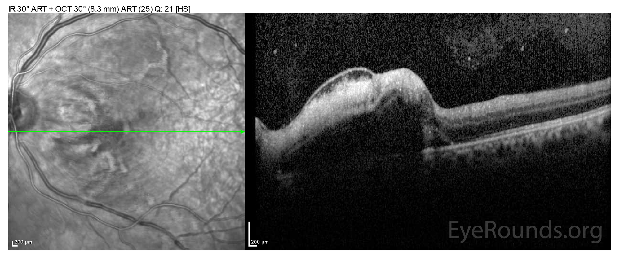
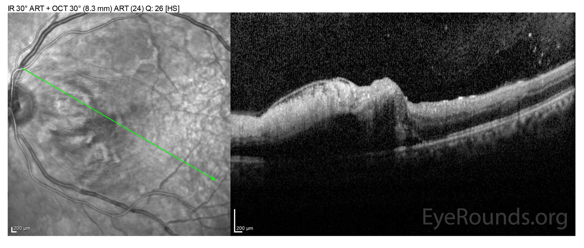
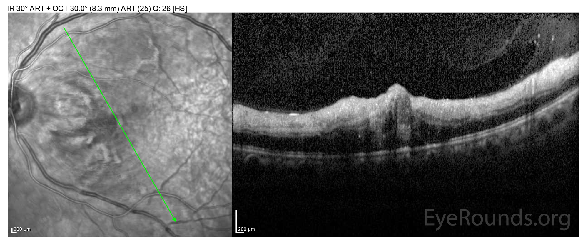
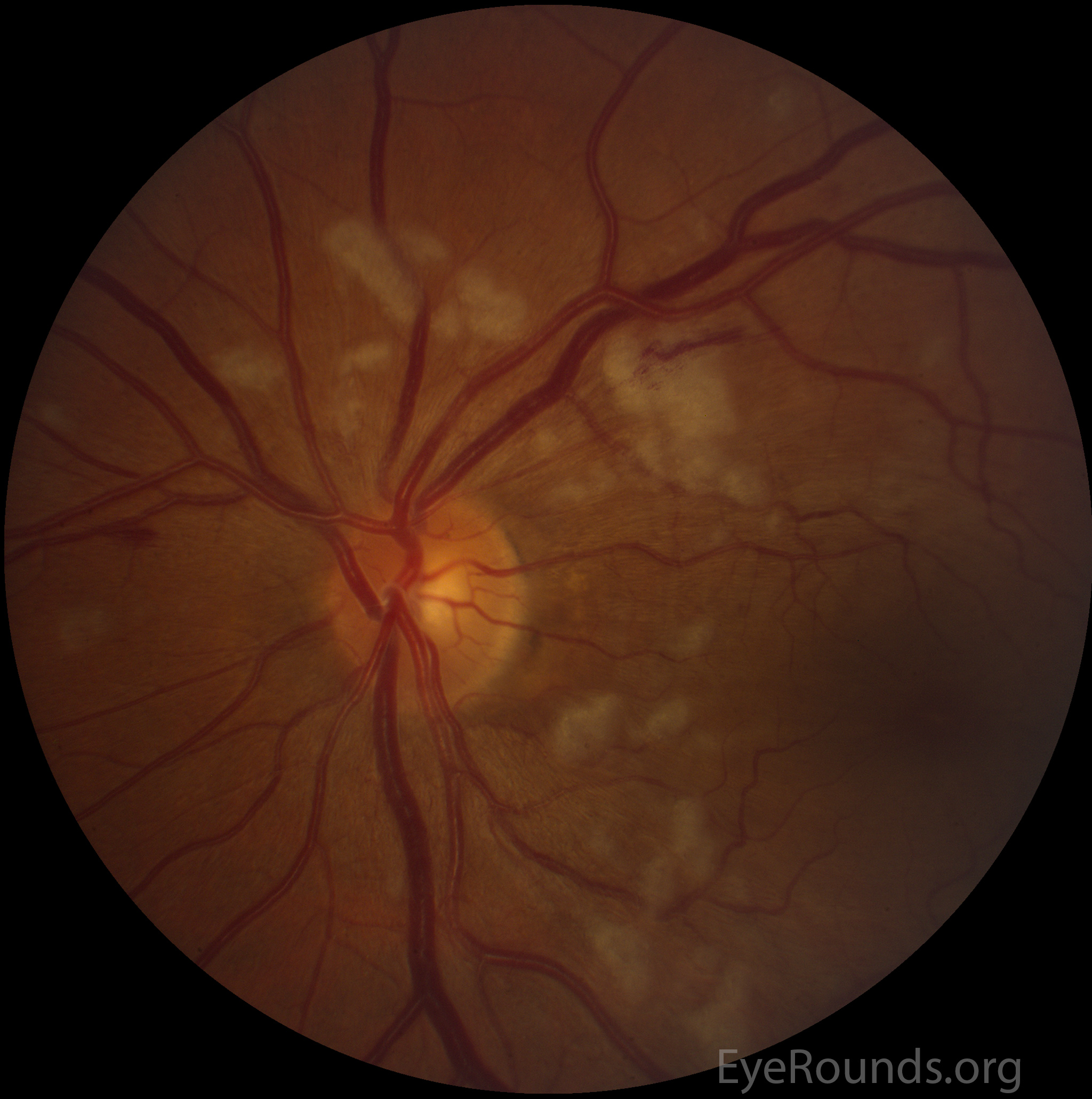
This is an impressive example of Purtscher's retinopathy following unrestrained motor vehicle accident. He suffered T5-T7 fracture, brain contusion, rib fracture, and mandibular fracture. He presented to clinic due to complaint of profound vision loss following his accident. On exam, he was noted to have Purtscher's retinopathy. Note the significant inner retinal edema on OCT and whitening, arteriolar hemorrhage, and edema of the fundus.
Purtscher's retinopathy typically follows trauma in the absence of direct trauma to the globe. Common causes are long bone fracture, chest compression injuries, air embolization, childbirth, as well as pancreatitis and connective tissue disorders. Clinical presentation is variable, with some patients presenting with profound vision loss and others with minimal changes. A relative afferent pupillary defect may or may not be present. Clinical findings are cotton wool spots, retinal hemorrhages, and Purtscher's flecken, which are areas of whitening in the inner retina between the retinal arterioles and venules. OCT displays hyperreflectivity of the inner retinal layers due to inner retinal edema. Prognosis is variable and largely depends on the extent of retinal involvement.
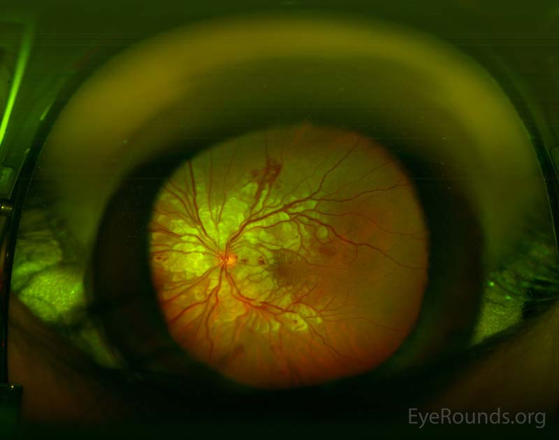
Purtscher retinopathy is the appearance of cotton wool spots, hemorrhages, and retinal edema located predominantly around the optic disc. It is caused by complement activation after acute crushing injuries to the head or thorax. Similar-appearing Purtscherlike retinopathy can be caused by a variety of etiologies including acute pancreatitis, chronic renal failure, autoimmune diseases, fat embolism, amniotic fluid embolism, retrobulbar anesthesia, and orbital steroid injection. Below are images of two separate patients with Purtscher retinopathy after compressive injuries to the chest.
Purtscher retinopathy is an occlusive microvasculopathy which classically results from head trauma, chest compression, or long bone fracture. This 25-year-old patient was involved in a high speed, roll-over motor vehicle crash and sustained both head trauma and chest compression injury resulting in a rib fracture. Visual acuity in the affected eye was 20/250 eccentrically. He was later lost to follow up. There also exists a related entity, Purtscher-like Retinopathy, which has the same retinal findings as Purtscher Retinopathy but occurs in the absence of trauma. Instead, Purtscher-like Retinopathy occurs in conditions such as acute pancreatitis, fat embolism syndrome, kidney failure, and childbirth. (1)

Ophthalmic Atlas Images by EyeRounds.org, The University of Iowa are licensed under a Creative Commons Attribution-NonCommercial-NoDerivs 3.0 Unported License.

Address
University of IowaLegal
Related Links