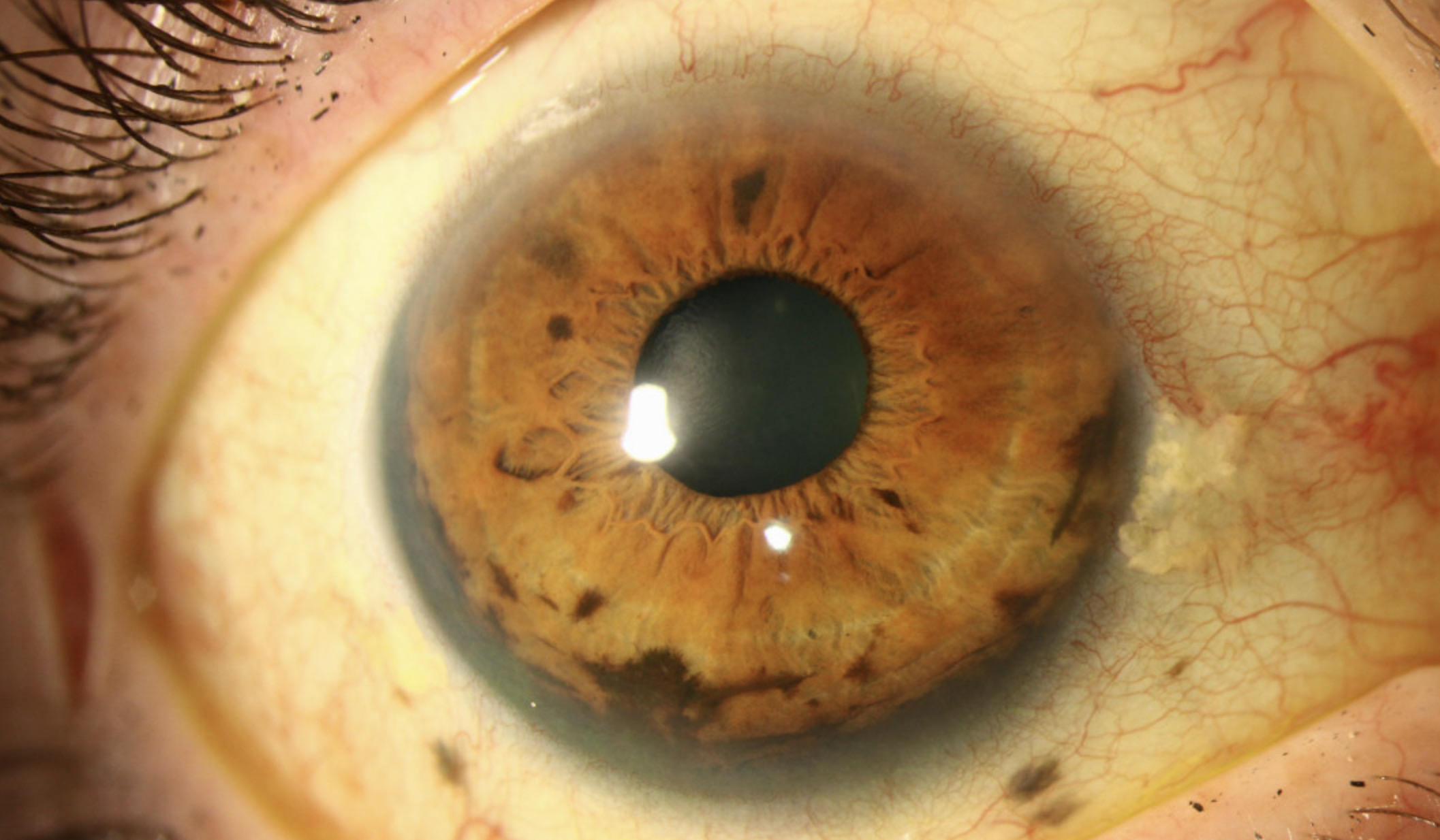
This is an 81-year-old gentleman who was referred to the University of Iowa glaucoma service for a history of acquired heterochromia, in addition to progressively elevated intraocular pressures. Upon presentation his anterior slit lamp exam showed a deep anterior chamber with 2+ pigmented cell. There was diffuse, velvet-like pigment on the iris in a circumferential fashion and also neovascularization. He had a posterior chamber intraocular lens. Gonioscopy revealed thick pigment overlying the entire iridocorneal angle for 360 degrees, causing his elevated IOP due to obstruction of the trabecular meshwork. Ocular echography revealed a ciliochoroidal lesion measuring 17mm at its base and 6mm in height. He was diagnosed with a ciliochoroidal melanoma leading to ring melanoma which prompted an immediate metastatic work up and subsequent enucleation.
A 58 year old woman presented for evaluation and treatment of an iris/ciliary body melanoma of the right eye. She initially noted scleral pigmentation which she initially thought was due to mascara. Upon presenting to her local ophthalmologist for worsening right eye pain, her IOP was elevated and she was found to have a pigmented lesion in the angle. Extensive, deep pigment was visualized 360 degrees in the angle on gonioscopy exam. There was also evidence of iris freckles and deep scleral pigment on slit lamp examination concerning for extraocular extension. The patient underwent high frequency anterior segment echography which demonstrated a lobular ring-like structure from 3-7 o'clock in the ciliary body with medium reflectivity which was consistent with an iridociliary melanoma. The patient ultimately underwent enucleation for local tumor control as plaque brachytherapy was unlikely to treat the extensive angle and extrascleral involvement.


Ophthalmic Atlas Images by EyeRounds.org, The University of Iowa are licensed under a Creative Commons Attribution-NonCommercial-NoDerivs 3.0 Unported License.