

Senile Calcific Plaque
Contributors: Kelly H. Yom, BA; Aaron M. Ricca, MD; Audrey C. Ko, MD
Photographer: Audrey C. Ko, MD
Posted June 26, 2018
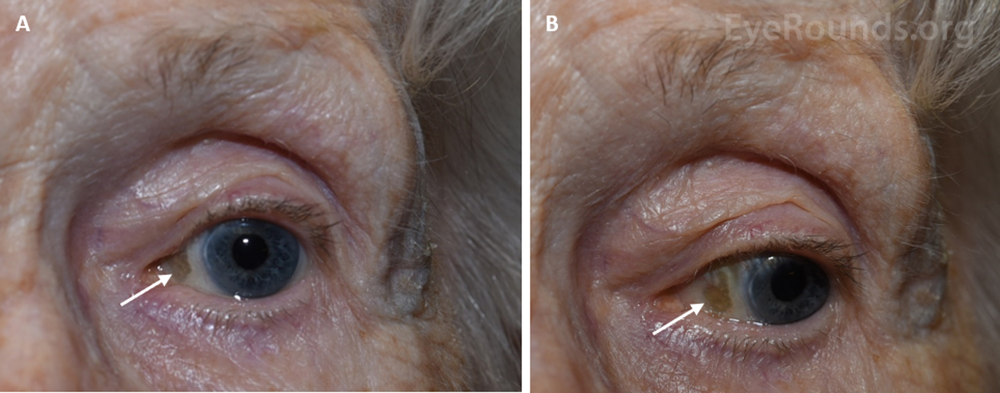
Figure 1. External photographs demonstrating a senile calcific plaque (arrows) found nasally in an 88-year-old woman and visible in primary (A) and lateral (B) gazes.
Senile calcific plaques are a relatively common incidental finding in the sclera of elderly patients. They are classically described as ovoid, well-demarcated, greyish lesions found just anterior to the insertion of the horizontal recti. Their appearance is attributed to age-related degeneration and subsequent calcification of the scleral hyaline [1]. This degeneration is not associated with structural weakening of the globe. Moreover, because they are usually asymptomatic, self-limiting, and not correlated with systematic disease, senile calcific plaques are rarely a cause for concern.
References
- Moseley I. Spots before the eyes: a prevalence and clinicoradiological study of senile scleral plaques. Clin Radiol. 2000; 55:198–206.

Ophthalmic Atlas Images by EyeRounds.org, The University of Iowa are licensed under a Creative Commons Attribution-NonCommercial-NoDerivs 3.0 Unported License.