Limbal Vernal Keratoconjunctivitis (VKC)
Contributor: Zachary Q. Mortensen, MD; Anthony T. Chung, MD; A. Tim Johnson, MD
Photographer: D. Brice Critser, CRA, OCT-C
Category:
- Cornea/External Eye Disease
Posted January 30, 2020
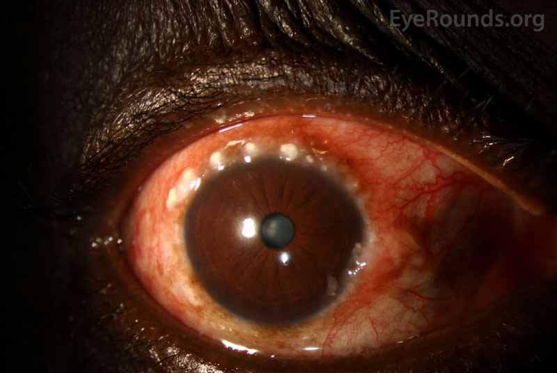
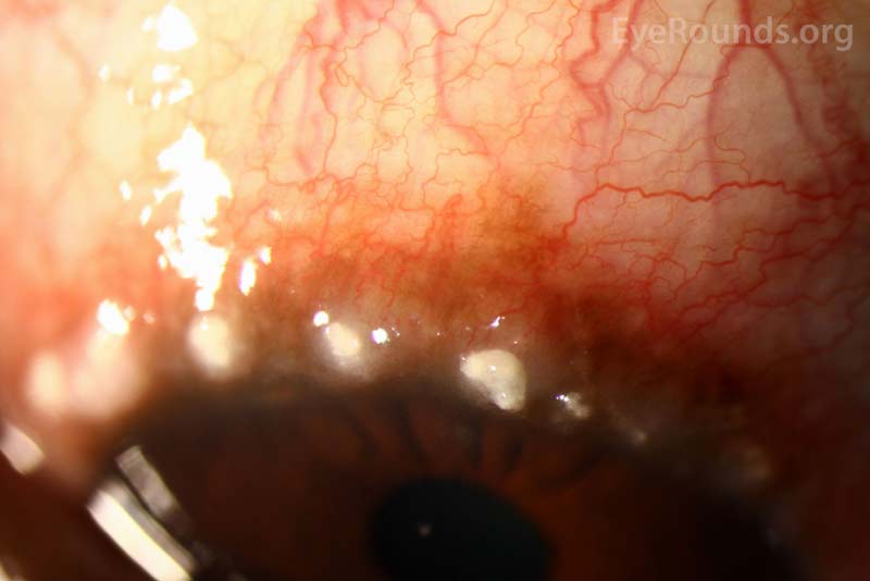
Limbal Horner-Trantas dots apparent on a 54-year-old African American male the setting of vernal keratoconjunctivitis.
Related: Vernal Keratoconjunctivitis: 8-year-old asthmatic male with reduced vision
Reference
- Koczman J, Oetting TA: Vernal Keratoconjunctivitis: 8-year-old asthmatic male with reduced vision. EyeRounds.org. June 25, 2007; Available from: http://www.EyeRounds.org/cases/70-Vernal-Keratoconjunctivitis-Atopic-Asthma.htm.
Limbal Vernal Keratoconjunctivitis
Contributor: David W Hayes, DO, US Military Flight Surgeon
Category: External Disease
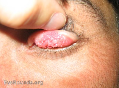
Severe VKC in a 17-year-old Afghani Male. Note the very large "cobblestone" papillae of the superior tarsal conjunctiva in both lids.
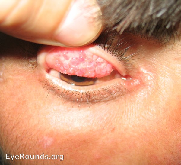
In this image, the concurrent bulbar conjunctival injection and limbal conjunctivitis is evident beneath the lid (see related EyeRounds case https://eyerounds.org/cases/70-Vernal-Keratoconjunctivitis-Atopic-Asthma.htm).
Limbal vernal keratoconjunctivitis (VKC)
Contributor: Jesse Vislisel, MD
Photographer: Stefani Karakas, CRA
Posted November 27, 2013
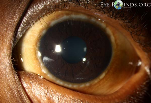
Vernal keratoconjunctivitis (VKC) is a seasonal disorder, predominantly seen in male children with a history of atopy, which results in inflammation of the cornea and conjunctiva. Limbal VKC is most common in children of African or Asian descent and may occur alone or in combination with palpebral VKC. Clinical features include Horner-Trantas dots (raised, white accumulations of eosinophils at the limbus), gelatinous limbal follicles, and copious amounts of mucoid discharge.
click image for higher resolution


Ophthalmic Atlas Images by EyeRounds.org, The University of Iowa are licensed under a Creative Commons Attribution-NonCommercial-NoDerivs 3.0 Unported License.

