

Adherent Leukoma
Contributor: William Charles Caccamise, Sr, MD, Retired Clinical Assistant Professor of Ophthalmology, University of Rochester School of Medicine and Dentistry
*Dr. Caccamise has very generously shared his images of patients taken while operating during the "eye season" in rural India as well as those from his private practice during the 1960's and 1970's. Many of his images are significant for their historical perspective and for techniques and conditions seen in settings in undeveloped areas.
Category: Cornea
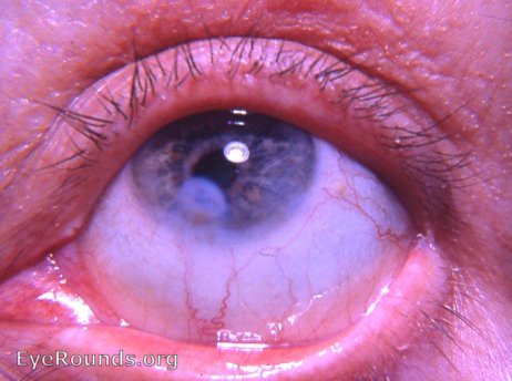
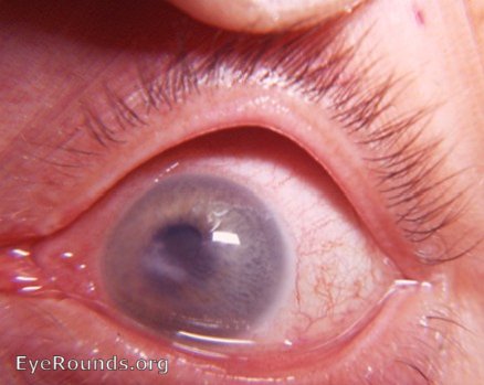
The history of this eye: A corneal ulcer formed. The ulcer produced a minute perforation of the cornea. The aqueous humor rushed towards the site of perforation and carried the iris with it.The iris plugged the perforation and became incorporated with the scar issue healing process. Notice the iris pigment that is incorporated in the corneal scar -evidence of iris involvement and thus, an adherent leukoma. Fortunately, this eye recovered from this potentially catastrophic situation. Other eyes might have gone on to staphyloma, glaucoma,endophthalmitis, panophthalmitis and / or phthisis bulbi from the same initial corneal ulcer.
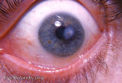
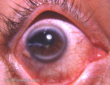
Adherent leukoma with aftercataract and with needling scar at limbus at 6 o'clock.
The patient had a perforating injury to his cornea. This injury was accompanied by an adherent leukoma and a secondary cataract. The cataract was later needled. The point of entrance of the discission knife can be seen as a scar at the limbus at 6 o'clock. An aftercataract occupies part of the pupil.
Adherent leukoma after knife wound
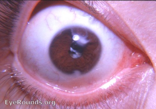
Adherent leukoma due to perforating corneal injury to only eye
At the age of 16 years, this 58-year-old patient lost his right eye due to a snowball accident. At the age of 58 years he presented with the pathology seen in the photo: the remaining eye had a perforating corneal scar with an adherent iris the cornea was vascularized and there was filamentary keratitis (seen with slit-lamp).
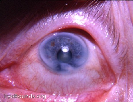
Scissors perforation of cornea: adherent leukoma, arcus senilis, and prominent pinguecula
The eye survived the injury insult with retention of very useful vision.


Ophthalmic Atlas Images by EyeRounds.org, The University of Iowa are licensed under a Creative Commons Attribution-NonCommercial-NoDerivs 3.0 Unported License.