This site uses tracking information. Visit our privacy policy. Click to agree to this policy and not see this again.

Ophthalmology and Visual Sciences

Basal cell carcinoma

Basal cell carcinoma

Category(ies): External Disease
Contributor: William Charles Caccamise, Sr, MD
Retired Clinical Assistant Professor of Ophthalmology, University of Rochester School of Medicine and Dentistry

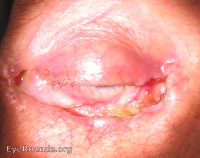
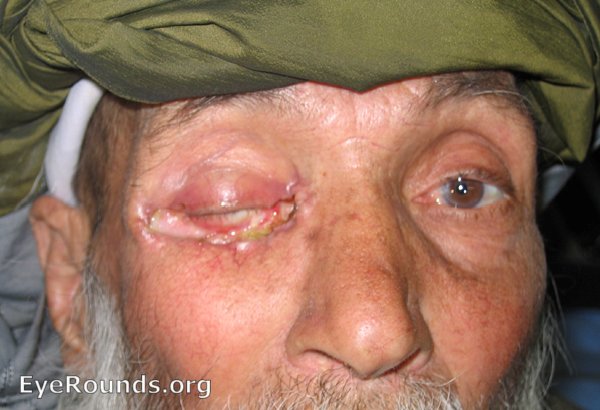
Dr. Caccamise has very generously shared his images of patients taken while operating during the "eye season" in rural India as well as those from his private practice during the 1960's and 1970's. Many of his images are significant for their historical perspective and for techniques and conditions seen in settings in undeveloped areas.


Figure 1: These first two photos show lesions that immediately suggests basal cell carcinoma unless proved otherwise by excisional biopsy. The pathologist's report inboth of these cases was " basal cell carcinoma " The lesions in these two photos is in the most common location for a basal cell carcinoma, i.e. the sun-exposed lower lid at the medial canthal area.
Basal Cell Carcinoma, Cheek
Figure 2: Because of changes in the appearance of this ugly lesion, an excisional biopsy was carried out - total removal followed by pathologist's diagnosis. The diagnosis was basal cell carcinoma with no excisional edge involvement.
Basal Cell Carcinoma, Near teat duct
Figure 3: This basal cell carcinoma is in a most demanding surgical location.The chances for having complications during and after surgery are great. The surgery in this case should be performed by an accomplished and experienced oculoplastic surgeon. The photo was taken by Dr. Caccamise in 1966.
Figure 4: This lesion had been apparent to the patient for 6 years. Excisional biopsy revealed basal cell carcinoma.
skin lesion has the typical characteristics of a basal cell carcinoma
Figure 5: The skin lesion has the typical characteristics of a basal cell carcinoma, i.e. the rolled edges with an umbilicated center. Nearby are some comedones.

Basal Cell Carcinoma, right lower lid

Basal Cell Carcinoma, right lower lid

Category(ies): Oculoplastics
Contributor: David W Hayes, DO US Military Flight Surgeon
alt text
Figure 6: Example of severe Basal Cell carcinoma with destruction of the entire right lower lid in a 61-year-old Afghani Male
alt text
Figure 7: Note the exposed bulbar conjunctiva and local destruction of tissue (reference Dr. O'Malley's case of a much smaller basal cell carcinoma in the lower lid and the surgical reconstruction plan).