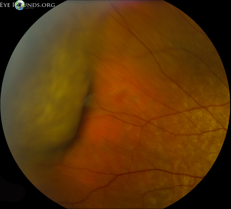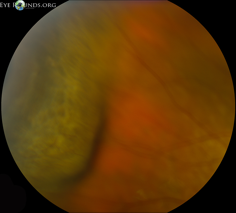


76-year-old male with hypertension and pseudophakia OU, presented to the retina clinic for cotton wool spots in the right eye. Several lab studies were done, in addition to carotid dopplers, all of which were unremarkable. An anterior chamber (AC) tap was done to rule out infectious etiologies (e.g. HSV, VZV). His vision in the right eye was 20/25 and IOP was 12. His slit lamp exam was unremarkable.
At one week follow-up, his vision was 20/40 and his IOP was 17. he said he had moderate discomfort immediately following his injection, but otherwise no other complaints. On exam he was found to have a localized hemorrhagic choroidal detachment in the superotemporal quadrant (see fundus photo). We presume from the history and sequence of events that his localized choroidal detachment resulted from his AC tap. This was observed and resolved over the following weeks.


Hemorrhagic choroidal detachments involve blood accumulation in the suprachoroidal space due to rupture of choroidal vessels.
Unlike serous choroidal effusions, which are typically painless, hemorrhagic choroidals have an abrupt onset often with severe pain.
Choroidal detachments are often managed conservatively. Often they resolve on their own.

Ophthalmic Atlas Images by EyeRounds.org, The University of Iowa are licensed under a Creative Commons Attribution-NonCommercial-NoDerivs 3.0 Unported License.