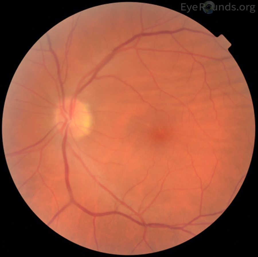
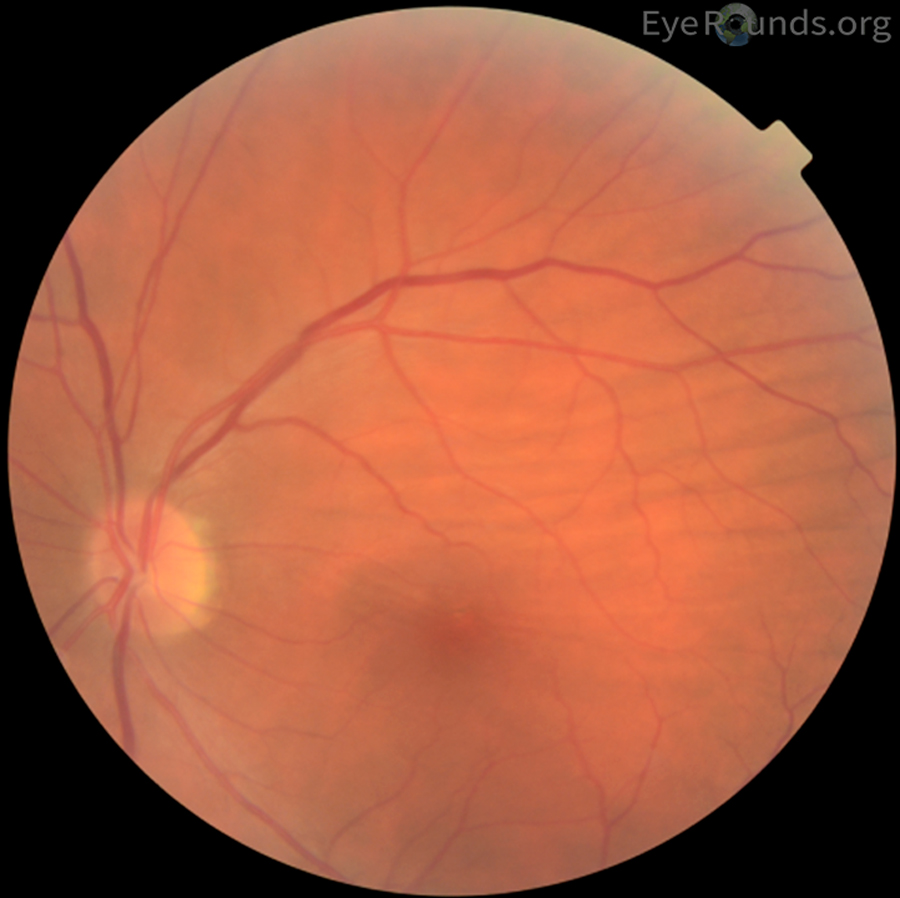
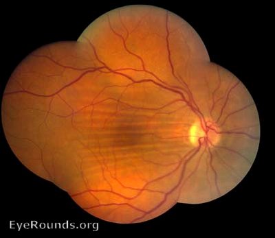
Choroidal folds occur when there is relative indentation of the inner surface of the sclera, leading to folds of the choroid and RPE or choroidal congestion. Clinically, they appear as parallel, alternating yellow and dark bands upon examination of the posterior pole. Choroidal folds may be present in hypotony, external globe compression, ocular or orbital inflammation, tumors, neovascular membranes, and papilledema.
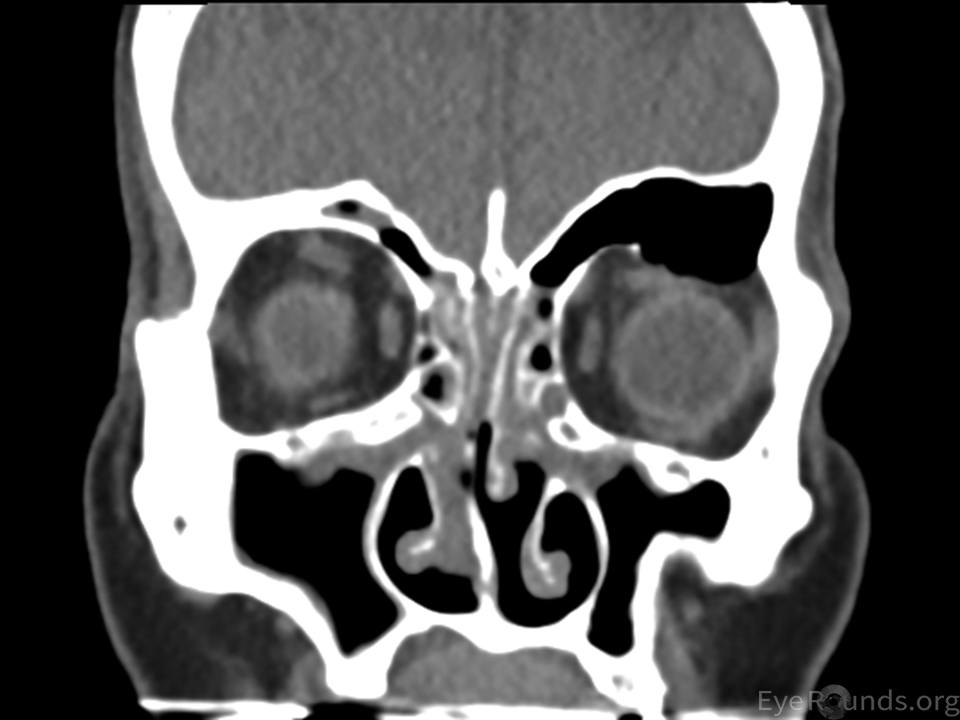
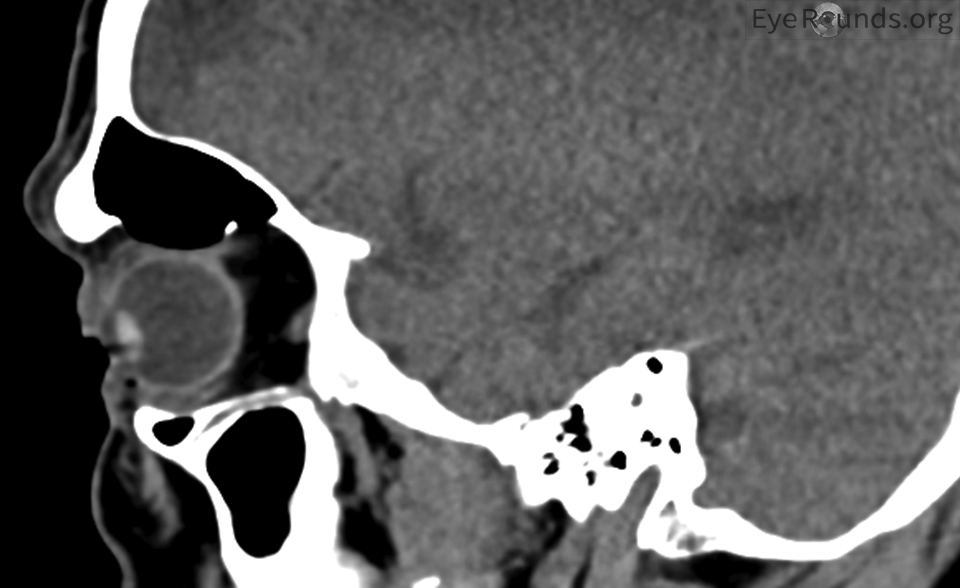
In this case, choroidal folds occurred secondary to external globe compression by air extending inferiorly into the orbit from the frontal sinus.

Ophthalmic Atlas Images by EyeRounds.org, The University of Iowa are licensed under a Creative Commons Attribution-NonCommercial-NoDerivs 3.0 Unported License.


Address
University of IowaLegal
Related Links