

EyeRounds Online Atlas of Ophthalmology
Contributor: William Charles Caccamise, Sr, MD, Retired Clinical Assistant Professor of Ophthalmology, University of Rochester School of Medicine and Dentistry
Expanded text by Brendan K. Penaluna, May 5, 2017
*Dr. Caccamise has very generously shared his images of patients taken while operating during the "eye season" in rural India as well as those from his private practice during the 1960's and 1970's. While the quality of his images are not up to today's higher web standards, many of his images are significant for their historical perspective and for techniques and conditions seen in settings in undeveloped areas.
Dendritic keratitis HSV - active
Active Dendritic Keratitis is often a clinical diagnosis and can be appreciated with fluorescein stain. New superficial ulcerations or expanding borders of the ulcer are signs of active infection.
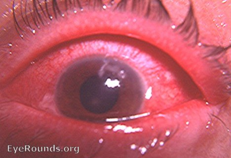
This is an example of acute hyperemic reaction to acute dendritic keratitis.

Reference
Biswell R. Chapter 6. Cornea. In: Riordan-Eva P, Cunningham ET, Jr. eds. Vaughan & Asbury's General Ophthalmology, 18e New York, NY: McGraw-Hill; 2011. http://accessmedicine.mhmedical.com/content.aspx?bookid=387§ionid=40229323. Accessed April 14, 2017.
originally posted 02-08-2008

Ophthalmic Atlas Images by EyeRounds.org, The University of Iowa are licensed under a Creative Commons Attribution-NonCommercial-NoDerivs 3.0 Unported License.