This site uses tracking information. Visit our privacy policy. Click to agree to this policy and not see this again.

Ophthalmology and Visual Sciences

Essential iris atrophy, iridocorneal endothelial (ICE) syndrome

Essential iris atrophy, iridocorneal endothelial (ICE) syndrome

Category(ies): Cornea, Iris
Contributor: Jesse Vislisel, MD
Photographer: Carol Chan, CRA

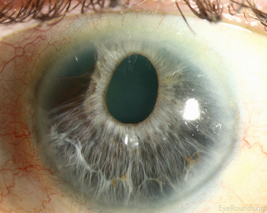
Essential iris atrophy is one of the subtypes of iridocorneal endothelial (ICE) syndrome. In this condition, an abnormally proliferating corneal endothelium grows across the trabecular meshwork and iris to form broad peripheral anterior synechiae (PAS), ectropion uveae, and iris stretch and melt holes. This photograph shows corectopia with a superotemporal iris melt hole in a patient with essential iris atrophy. The patient also had extensive PAS on gonioscopy.


Essential Iris Atrophy

Essential Iris Atrophy

Category(ies): Glaucoma / Iris
Contributor: Mahsaw Mansoor, MD
Photographer: Jody Troyer, CRA
Posted: 06/04/2024


Essential iris atrophy, iridocorneal endothelial (ICE) syndrome

Essential iris atrophy, iridocorneal endothelial (ICE) syndrome

Category(ies): Glaucoma / Iris
Contributor: Emma M. Hartness, MD; Patrick Donegan, MD; Marc Toeteberg-Harms, MD
Photographer: Jennifer A. Lynch
Posted: 8/3/2026

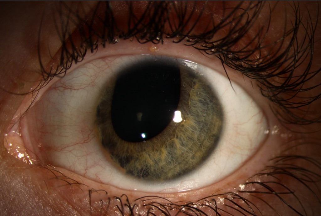
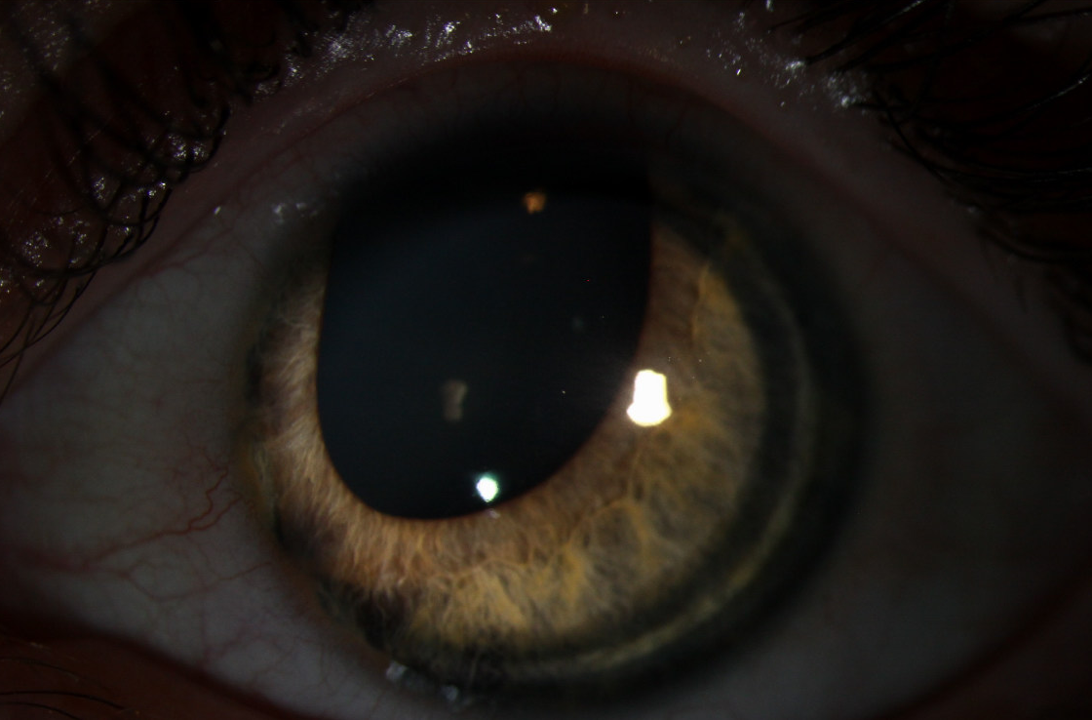
A 54-year-old woman was originally referred to the glaucoma service by the optometry clinic for right pupil dyscoria. Examination originally showed a mild beaten-bronze appearance of the corneal endothelium, peripheral anterior synechiae extending past Schwalbe line, and a slight nasal peaking of the iris which progressed over several years into a horizontal ovoid dyscoria. She developed intraocular hypertension and was treated with one drop of timolol in the morning in the right eye.

Slit lamp photograph of the right eye showing iris atrophy with an ovoid, horizontal corectopia
Figure 1. Slit lamp photograph of the right eye showing iris atrophy with an ovoid, horizontal corectopia.
 Slit lamp gonioscopy photograph of the right eye showing temporal high peripheral anterior synechiae with ectropion uveae covering the angle
Figure 2A. Slit lamp gonioscopy photograph of the right eye showing temporal high peripheral anterior synechiae with ectropion uveae covering the angle.
Slit lamp gonioscopy photograph of the right eye showing nasal high peripheral anterior synechiae covering the angle with mild ectropion uveae.
Figure 2B. Slit lamp gonioscopy photograph of the right eye showing nasal high peripheral anterior synechiae covering the angle with mild ectropion uveae.
Slit lamp photograph of the right eye demonstrating dilation of the pupil with horizontal dyscoria.
Figure 3. Slit lamp photograph of the right eye demonstrating dilation of the pupil with horizontal dyscoria.