

Familial corneal dystrophy
Contributor: William Charles Caccamise, Sr, MD, Retired Clinical Assistant Professor of Ophthalmology, University of Rochester School of Medicine and Dentistry
*Dr. Caccamise has very generously shared his images of patients taken while operating during the "eye season" in rural India as well as those from his private practice during the 1960's and 1970's. Many of his images are significant for their historical perspective and for techniques and conditions seen in settings in undeveloped areas.
Category: Cornea
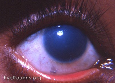
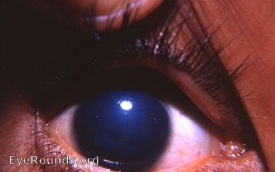
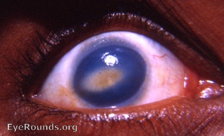
Several siblings - males and 1 female - were followed periodically for several years at the Eye Clinic. Initially, the involvement seemed to be confined to the epithelium. Later, the lesions seemed to be a bit deeper. In addition, fatty infiltration occurred. Several male members of the family have the same corneal dystrophy.
Initial photos taken in 1964
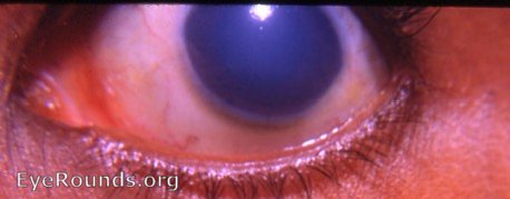
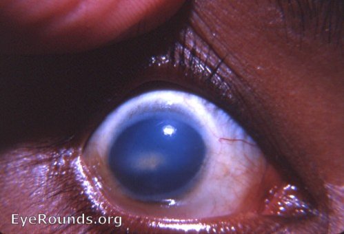
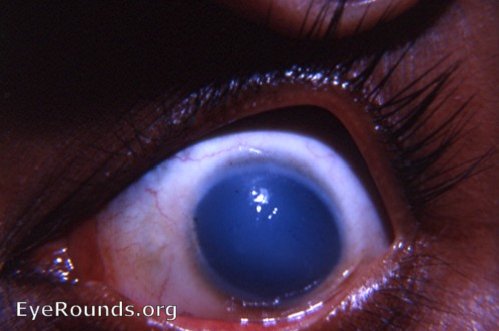
This family was followed by Dr. Caccamise for a number of years - from an initial slight epithelial involvement to a later fatty infiltration. Photo of an older brother's eye was taken in 1966. In previous years, that eye had demonstrated the same appearance demonstrated above. In the photos belowfatty changes have occurred in the older brother's cornea.

Ophthalmic Atlas Images by EyeRounds.org, The University of Iowa are licensed under a Creative Commons Attribution-NonCommercial-NoDerivs 3.0 Unported License.