88-year-old male referred for evaluation of an epiretinal membrane and lamellar hole in the right eye. Patient had a history of a stroke with secondary visual field defects (quadrantanopsia). No acute vision changes. No metamorphopsia. No pain or irritation. No other complaints.
• BCVA cc: OD 20/50; OS 20/25
• IOP: 19 and 21
• SLE: PCIOL OU
Discussed surgery versus observation. The patient elected for observation of his epiretinal membrane and lamellar hole.
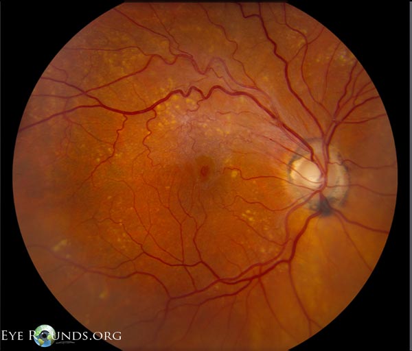
Figure 1: OD: clear media; optic nerve cupping with peripapillary pigment; multiple intermediate and large drusen throughout macula and periphery; small lamellar hole at the fovea center and an epiretinal membrane worse between the fovea and superior arcades.
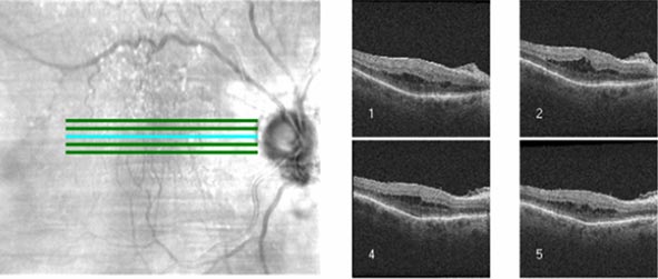
•Seen on slit lamp biomicroscopy as a round central inner retinal defect without thickening, cystic change, or subretinal fluid.
•An overlying operculum is often seen (but not present in this case).
•Mechanism is secondary to vitreofoveal separation with loss of the inner retinal layers, but with a preserved and intact outer photoreceptor layer.
•Vision is usually good (20/20-20/30) and patients are often asymptomatic, when no other confounding variables such as an epiretinal membrane are present.
•Fluorescein angiography shows no abnormal fluorescence.
University of Iowa
Roy J. and Lucille A. Carver College of Medicine
Department of Ophthalmology and Visual Sciences
200 Hawkins Drive
Iowa City, IA 52242
University of Iowa
Roy J. and Lucille A. Carver College of Medicine
Department of Ophthalmology and Visual Sciences
200 Hawkins Drive
Iowa City, IA 52242