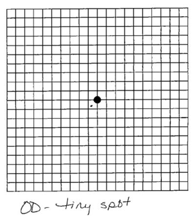
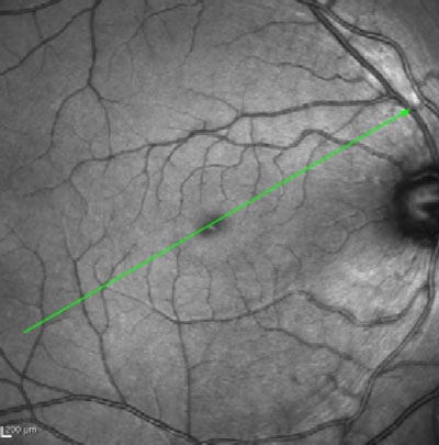
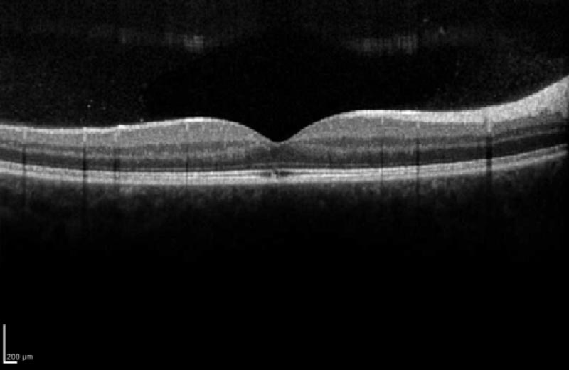
This is a 16-year-old male who suffered a laser pointer injury to his right eye and subsequently found that he had a small central area of "blackness" that made it difficult to read. On fundus exam, the right eye showed a small, depigmented, circular defect just temporal to the center of the fovea. OCT showed a small cavitation at this location with IS/OS disruption. The Amsler grid was normal for the left eye but demonstrated a small central scotoma in the right eye. The exam was otherwise normal including 20/20 vision OU.
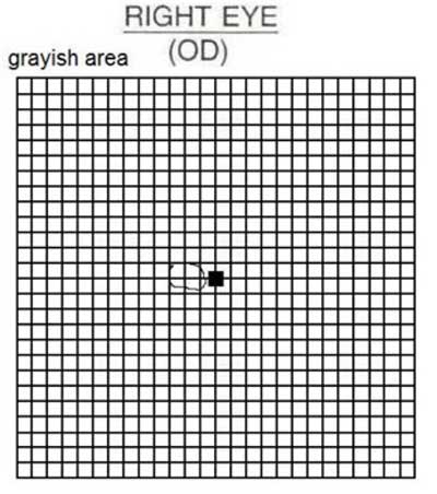
On one month follow-up, Amsler grid testing demonstrated a reduced scotoma size, and the patient reported that he notices the deficit less. The visible lesion near the fovea had healed somewhat and appeared smaller on OCT and fundus exam.

Ophthalmic Atlas Images by EyeRounds.org, The University of Iowa are licensed under a Creative Commons Attribution-NonCommercial-NoDerivs 3.0 Unported License.


Address
University of IowaLegal
Related Links

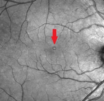
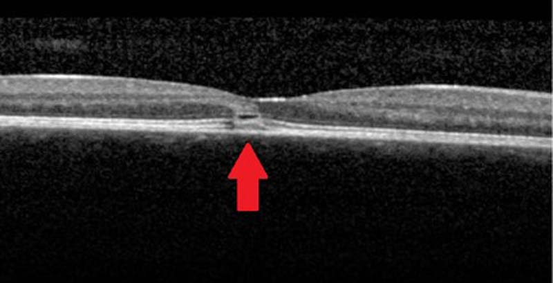 2 days after injury