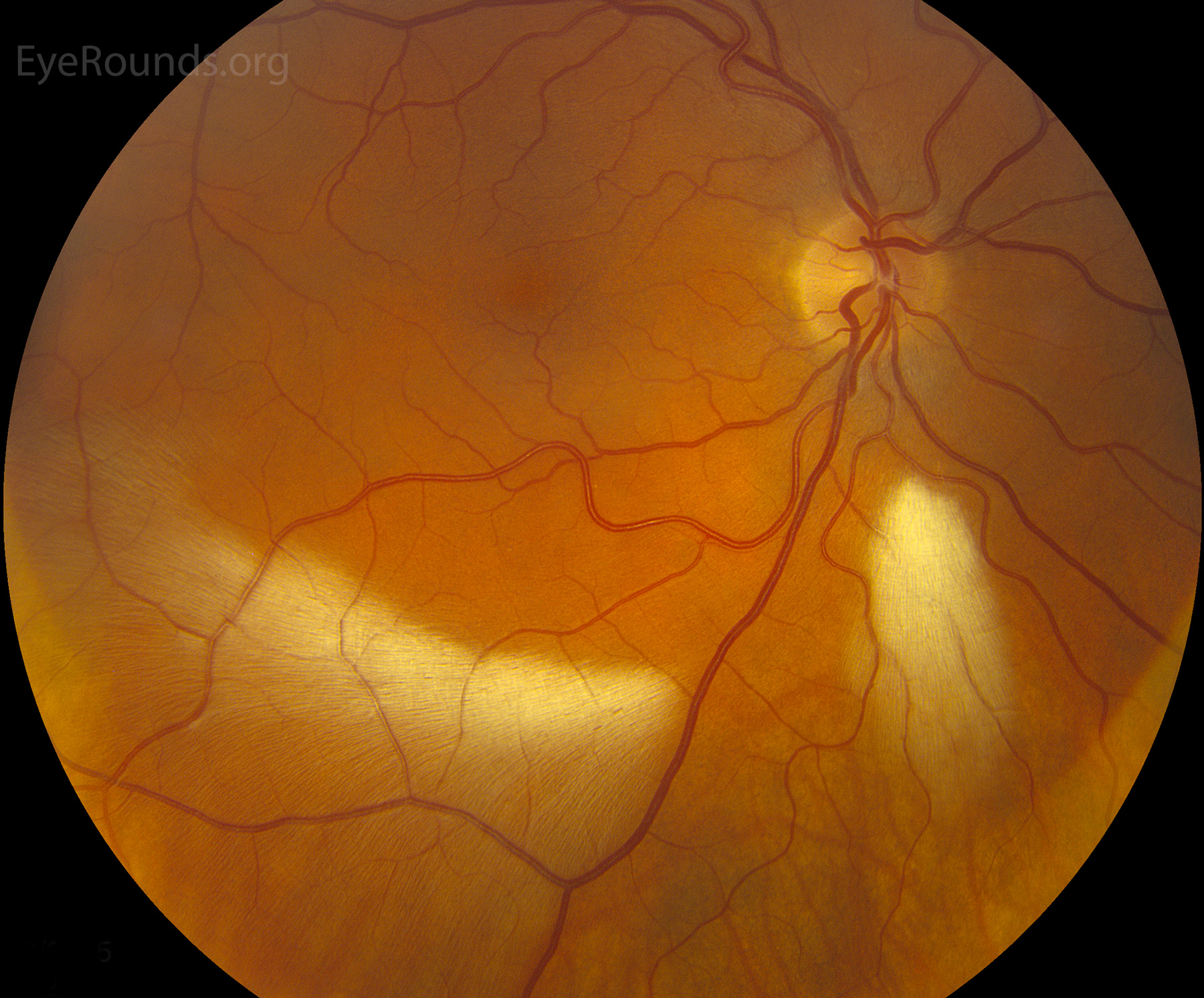
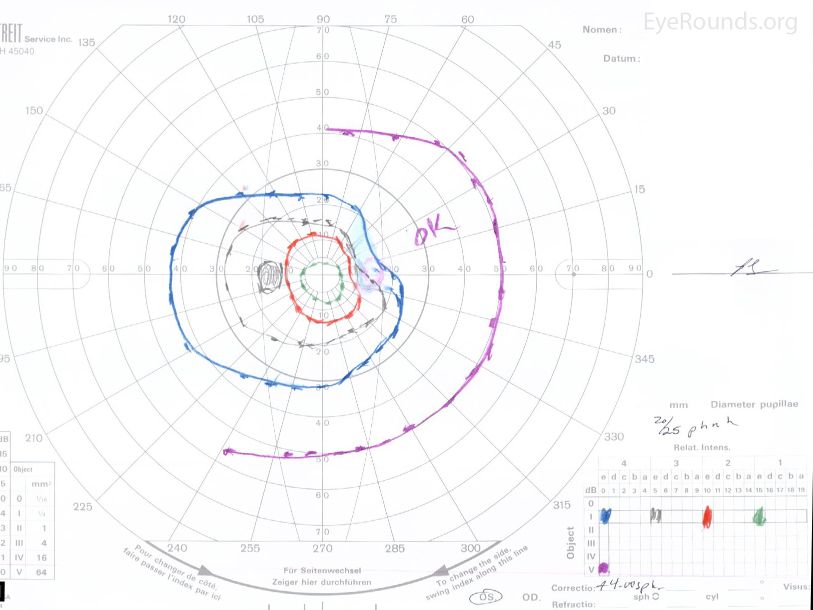
This is an example of a prominent myelinated nerve fiber layer. These can have corresponding visual field defects as seen here on Goldmann perimetry with depression of the I4e, I3e, and I2e isopters in the region of the myelination. This is an unusual case as it is located along the horizontal raphe; typically, the myelin is peripapillary in location.

Ophthalmic Atlas Images by EyeRounds.org, The University of Iowa are licensed under a Creative Commons Attribution-NonCommercial-NoDerivs 3.0 Unported License.

Address
University of IowaLegal
Related Links