

EyeRounds Online Atlas of Ophthalmology
Contributor: William Charles Caccamise, Sr, MD, Retired Clinical Assistant Professor of Ophthalmology, University of Rochester School of Medicine and Dentistry
*Dr. Caccamise has very generously shared his images of patients taken while operating during the "eye season" in rural India as well as those from his private practice during the 1960's and 1970's. Many of his images are significant for their historical perspective and for techniques and conditions seen in settings in undeveloped areas.
Category: Pediatrics
Nanophthalmos with cataract
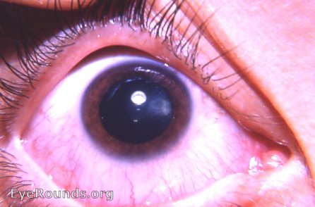
Under full mydriasis, a peripheral cataractous change is seen at 3 o'clock OD. This should be followed to determine any future activity. It may congenital and stationary. The second photo shows both eyes of this patient.The OD has nanopthalmos.
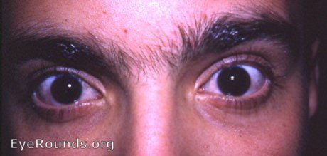
both eyes of the same patient as above. Right eye is the eye with nanophthalmos. Careful measurement will reveal that the right eye is smaller than the left eye but appears normal. When the eye is smaller than normal but is otherwise normal, nanophthalmos is said to exist. Some use microphthalmos synonymously with nanophthalmos. Dr. Caccamise prefers to use micropthalmos for the small eye that has additional apparent abnormalities, e.g. microcornea with opacification, microcornea with coloboma of the iris, microcornea with cataract, etc.

Ophthalmic Atlas Images by EyeRounds.org, The University of Iowa are licensed under a Creative Commons Attribution-NonCommercial-NoDerivs 3.0 Unported License.