This site uses tracking information. Visit our privacy policy. Click to agree to this policy and not see this again.

Ophthalmology and Visual Sciences

Ocular toxoplasmosis

Ocular toxoplasmosis

Category(ies): Retina, Vitreous, Uveitis
Contributor: Jesse Vislisel, MD
Photographer: Brice Critser, CRA (fig 1), Stefani Karakas, CRA(fig 2)

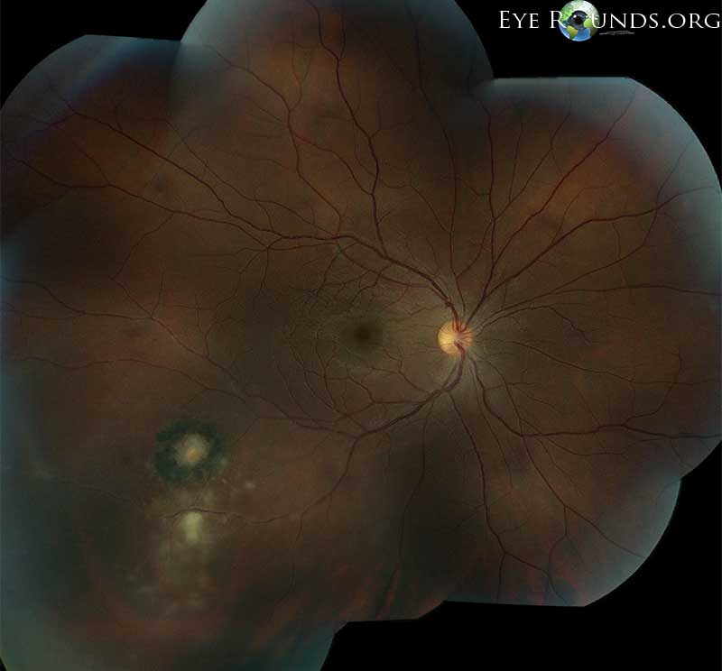
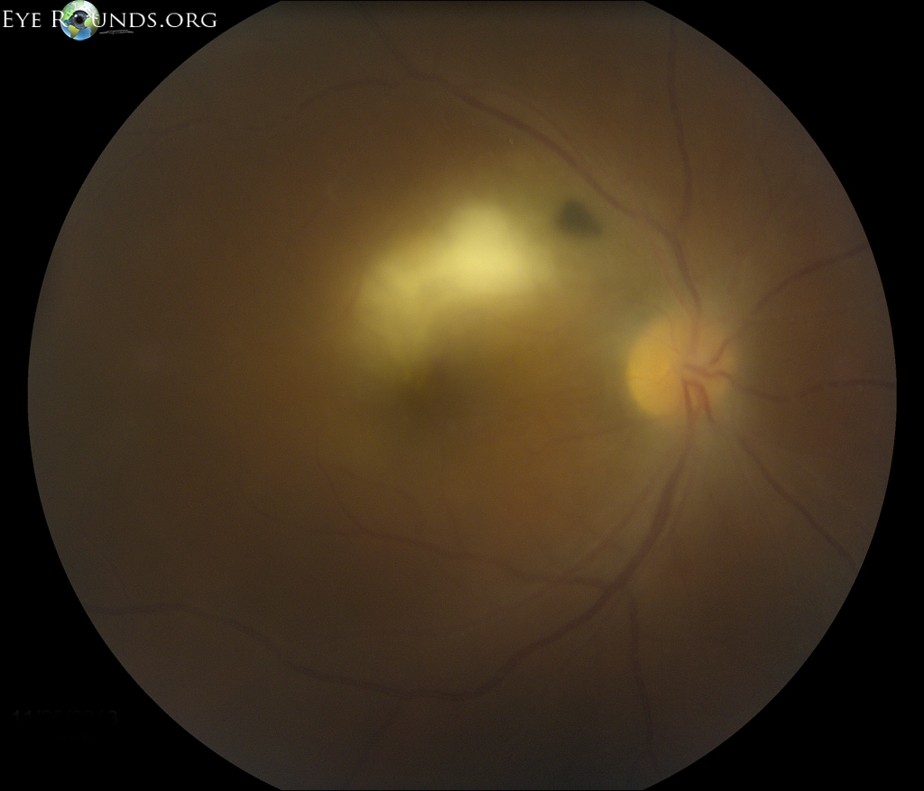
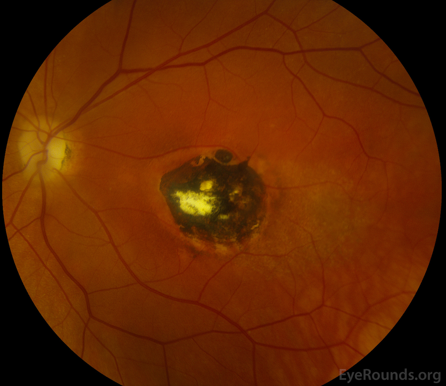
Toxoplasmosis is the most common cause of posterior uveitis. Active lesions have a classic "headlight in fog" appearance with a focal, white, fluffy lesion adjacent to an old scar visible through the associated granulomatous uveitis and vitritis as seen in Figure 1. Inactive lesions appear as a chorioretinal scar in the posterior pole, often within the macula as seen in Figure 2.

fundus photo showing active lesions
Active lesions have a classic "headlight in fog" appearance with a focal, white, fluffy lesion adjacent to an old scar visible through the associated granulomatous uveitis and vitritis as seen in Figure 1
fundus photo showing inactive lesions
Inactive lesions appear as a chorioretinal scar in the posterior pole, often within the macula as seen in Figure 2.

Contributor: Lauren E. Hock, MD
Photographer: Meghan Menzel
fundus photo showing inactive lesions
Congenital toxoplasmosis with macular chorioretinal scar seen in a 69-year-old woman who presented with severe primary open angle glaucoma of both eyes. Vision had been limited to CF @2 ft in the affected eye since birth.