This site uses tracking information. Visit our privacy policy. Click to agree to this policy and not see this again.

Ophthalmology and Visual Sciences

Peri-Vascular Lattice Degeneration

Peri-Vascular Lattice Degeneration

Category(ies): Retina, Vitreous
Contributor: Eric Chin, MD
Photographer: Stefani Karakas

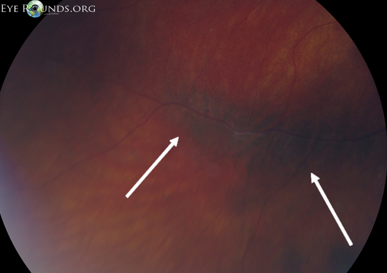
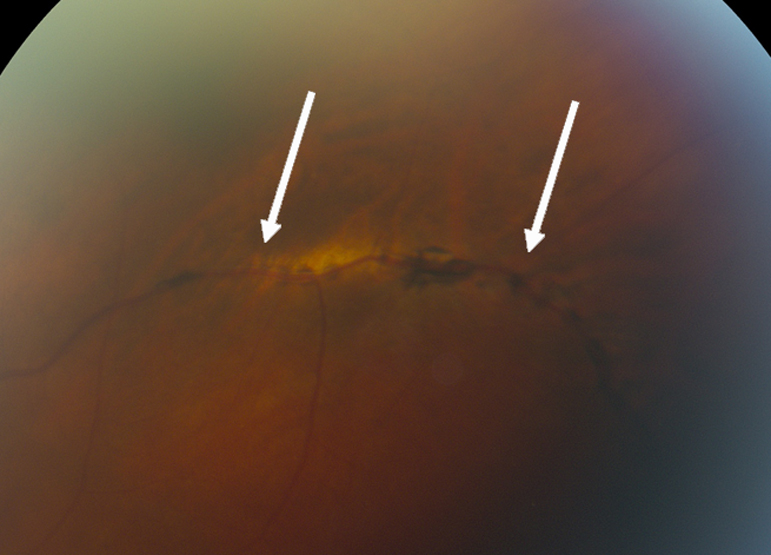
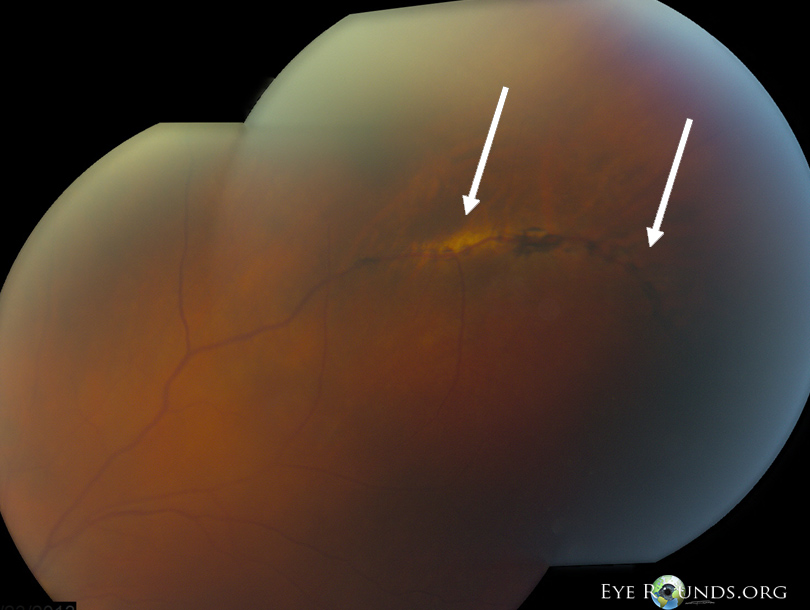
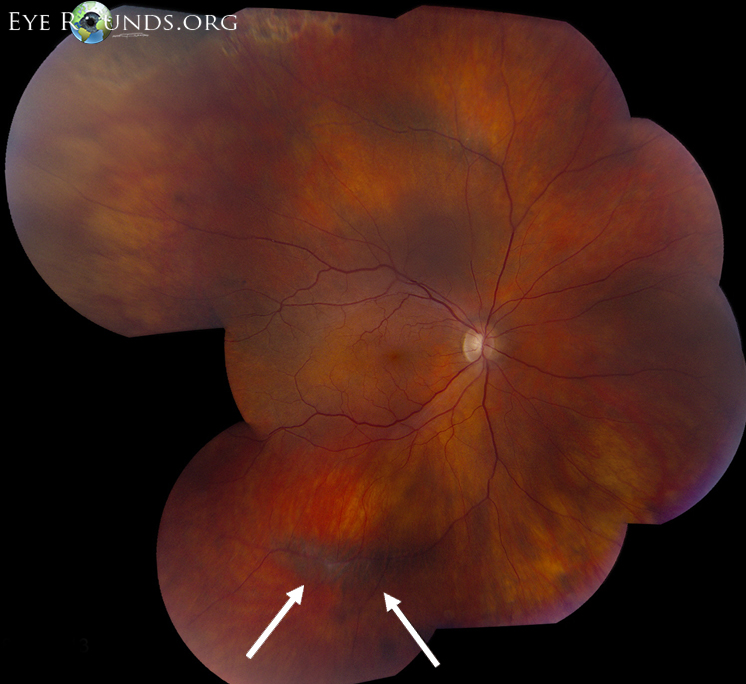
74-year-old female who underwent recent epiretinal membrane peel in the right eye. Intraoperatively, she was found to have several small retinal tears in the periphery which were lasered for prophylaxis.

  • • BCVA: OD 20/20
  • • IOP: normal
  • • SLE: unremarkable
  • • DFE: OD next slide
 OD: radial perivascular lattice with sclerotic vessels and hyperplastic retinal pigment epithelium
Figure 1: OD: radial perivascular lattice with sclerotic vessels and hyperplastic retinal pigment epithelium
OD: higher magnification of radial perivascular lattice
Figure 2: OD: higher magnification of radial perivascular lattice
OS: radial perivascular lattice
Figure 3: OS: radial perivascular lattice
OS: radial perivascular lattice
Figure 4: OS: radial perivascular lattice

Lattice Degeneration

  • Histopathologically represents localized areas of inner retinal atrophy with loss of the internal limiting membrane, adherent vitreous to the edges of the lesion with an overlying pocket of liquefied vitreous, and sclerosis of the remaining retinal vessels.
  • Lattice degeneration is seen in approximately 10% of the population, but rarely leads to rhegmatogenous retinal detachments.
  • However, lattice degeneration is present in 20-40% of rhegmatogenous detachments.

Radial Perivascular Lattice Degeneration

  • Lattice lesions with retinal thinning and pigmentary disturbances along retinal vessels may be referred to as radial perivascular chorioretinal degeneration.
  • Typically seen more posteriorly along retinal vessels in comparison to traditional peripheral lattice degeneration.
  • May be an incidental finding, as in this case, or seen with hereditary vitreoretinal degenerations such as Stickler syndrome in which there is a high risk for rhegmatogenous retinal detachment.
References:
  1. Byer NE. Long-term natural history of lattice degeneration of the retina. Ophthalmology 1989;96:1396-1402.
  2. Lewis H. Peripheral retinal degenerations and the risk of retinal detachment. Am J Ophthalmol 2003;136:155-160.

Image Permissions:
Creative Commons

Ophthalmic Atlas Images by EyeRounds.org, The University of Iowa are licensed under a Creative Commons Attribution-NonCommercial-NoDerivs 3.0 Unported License.