

EyeRounds Online Atlas of Ophthalmology
Contributor: William Charles Caccamise, Sr, MD, Retired Clinical Assistant Professor of Ophthalmology, University of Rochester School of Medicine and Dentistry
*Dr. Caccamise has very generously shared his images of patients taken while operating during the "eye season" in rural India as well as those from his private practice during the 1960's and 1970's. Many of his images are significant for their historical perspective and for techniques and conditions seen in settings in undeveloped areas.
Category: External Disease
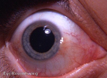
Pinguecula
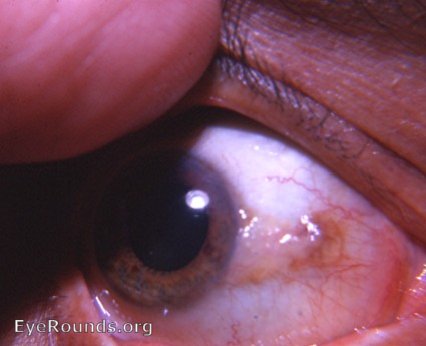
The photo shows an unusually large pinguecula involving the nasal bulbar conjunctiva. Although it appears to consist of fat, it actually consists of conjunctival tissue involved with elastic tissue degeneration. Histologically, it is similar to a pterygium without corneal involvement.
Pinguecula? Starting point for a pterygium?
Since the conjunctival component of a pterygium is identical to a pinguecula, it has been claimed by some pathologists that the pterygium originates from a pinguecula.
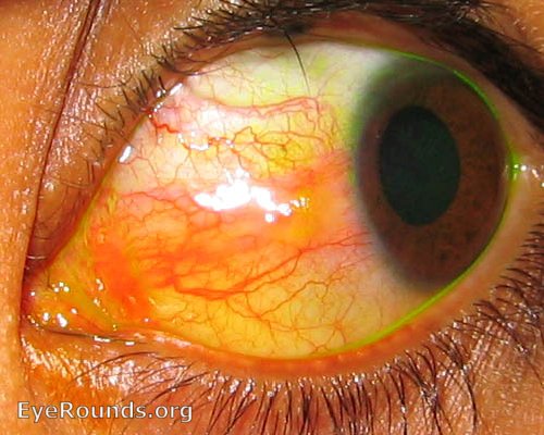
Pinguecula - inflamed
Contributor: Andrew Doan, MD, PhD, University of Iowa
Category: External Disease

Ophthalmic Atlas Images by EyeRounds.org, The University of Iowa are licensed under a Creative Commons Attribution-NonCommercial-NoDerivs 3.0 Unported License.