

EyeRounds Online Atlas of Ophthalmology
Contributor: William Charles Caccamise, Sr, MD, Retired Clinical Assistant Professor of Ophthalmology, University of Rochester School of Medicine and Dentistry
*Dr. Caccamise has very generously shared his images of patients taken while operating during the "eye season" in rural India as well as those from his private practice during the 1960's and 1970's. Many of his images are significant for their historical perspective and for techniques and conditions seen in settings in undeveloped areas.
Category: Cataract
Posterior subcapsular cataract
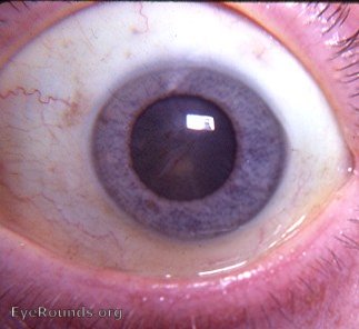
Under full mydriasis, the posterior subcapsular cataract is best diagnosed by slit-lamp examination. Ophthalmoscopic examination will also lead to a correct diagnosis. In the photograph, it is not possible for the viewer to localize the cataractous changes to the posterior subcapsular zone of the lens. In Dr. Caccamise's practice in the USA, primary (idiopathic) posterior subcapsular cataract was a frequent reason for cataract surgery in those patients beyween 40 and 60 years of age. Secondary posterior subcapsular cataract is a common result of intraocular pathology, systemic disease or medication - especially oral steroid therapy.

Ophthalmic Atlas Images by EyeRounds.org, The University of Iowa are licensed under a Creative Commons Attribution-NonCommercial-NoDerivs 3.0 Unported License.