

Viral keratoconjunctivitis
EyeRounds Online Atlas of Ophthalmology
Contributor: William Charles Caccamise, Sr, MD, Retired Clinical Assistant Professor of Ophthalmology, University of Rochester School of Medicine and Dentistry
*Dr. Caccamise has very generously shared his images of patients taken while operating during the "eye season" in rural India as well as those from his private practice during the 1960's and 1970's. Many of his images are significant for their historical perspective and for techniques and conditions seen in settings in undeveloped areas.
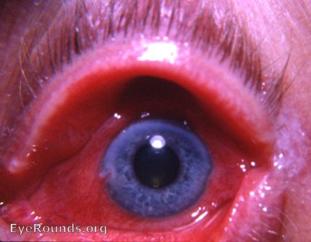
Photo of second case of this highly contagious disease. Limbus lesions are seen at 9 and 3 o'clock. With the slit-lamp, corneal infiltrates are readily evident in the pupillary area. They are only vaguely visible in the photo. Refer to the discussion that accompanies Dr. Caccamise's initial photo of this disease. Because of the contagious nature of this disease, all office followup visits should be scheduled after all other patients for the day have left the office. Like well-recognized nosocomial infections, the ophthalmologist's office has frequently been the source of epidemics of viral keratoconjunctivitis. Disposable instruments and repeated handwashing must be emphasized. There should be a minimum ofoffice visits by these patients.


Ophthalmic Atlas Images by EyeRounds.org, The University of Iowa are licensed under a Creative Commons Attribution-NonCommercial-NoDerivs 3.0 Unported License.