
Chief Complaint: Excess tearing
History of Present Illness: A 32-year-old gentleman presents with a 10-year history of excess tearing in both eyes. There has also been some mucous-like yellowish discharge. He states that the excess tearing henders his vision.
Past Ocular History: None
Allergies: No known drug allergies
Family History: Non-contributory
Social History: Non-contributory
Review of systems: Negative on 12 -point review of systems
Pupils: 4mm → 2mm, brisk, equal OU, no RAPD OU
Extraocular movements: Full, orthotropic OU
Confrontation visual field: Decreased peripheral visual field superiorly OU
Hertel exophthalmometry: 6mm OD, 5mm OS, base 97mm
 |
 |
 |
Dilated Fundus Exam: Normal disc, macula, vasculature and periphery OU
 |
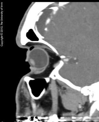 |
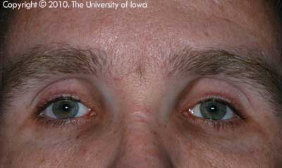
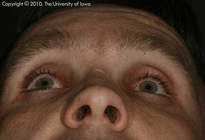
Diagnosis: Acquired enophthalmos consistent with silent brain syndrome
Bilateral secondary enophthalmos after ventriculo-peritoneal shunting (VPS) for hydrocephalus at a young age was first described in literature in 1996 by Meyer and colleagues. They described three patients with progressive severe bilateral enophthalmos who each had congenital hydrocephalus treated with VP shunts (Meyer, 1996).
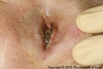
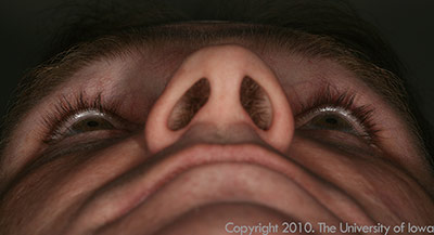
Due to the severe lack of lid apposition to the globe, these patients, initially, present to ophthalmologists seeking treatment for dry, irritative ocular symptoms. This may include a foreign body sensation, tearing, epiphora, or less likely, decreased vision. Some patients do state a chief complaint of their eyes "sinking into the sockets." In addition, there can be entrapment of air underneath the eyelids (Figure 5). Most patients have suffered from progressive worsening of these symptoms for multiple years prior to presenting to an ophthalmologist.
These patients, like our patient, usually have a previous history of hydrocephalus requiring a VPS early in life.
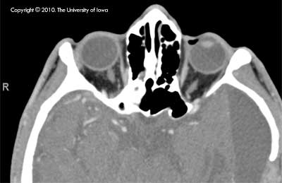
In 2008, Cruz et al. were the first to propose that VP shunting early in life can cause a reduction in size of the anterior cranial fossa, which, in turn, alters the structure of the orbit and leads to the development of enophthalmos. In young children, the thin floor of the frontal cranial fossa or the orbital roof is thought to be less calcified and, therefore, more susceptible to remodeling. These bones become progressively bowed superiorly over time as the intracranial pressure decreases (Cruz, 2008).
Bernadini et al. reported two patients with similar findings and labeled the disease as the "silent brain syndrome" (Bernadini, 2009). The silent brain syndrome is the counterpart to the "silent sinus syndrome," (Please see the EyeRounds case, "Silent sinus syndrome")
Patients with silent brain syndrome can be treated with orbital volume augmentation since the enlarged orbital cavity is responsible for the enophthalmos, as well as the consequent absence of lid apposition and air entrapment. Surgically, an appropriately-sized implant can be placed to bridge the bowed orbital roof. If an implant is not used, lid malposition repair can be performed. However, this option is usually not as successful in resolving the symptoms as orbital roof implants. Post-operatively, the patients may need to be evaluated for strabismus surgery. Our patient underwent bilateral orbital implants which improved his enophthalmos and his presenting ocular symptoms.
 |
Epidemiology:
|
Signs:
|
Symptoms:
|
Treatment:
|
Bernadini FP, Rose GE, Cruz AAV, Priolo E. Gross Enophthalmos After Cerebrospinal Fluid Shunting for Childhood Hydrocephalus: The "Silent Brain Syndrome". Ophthal Plast Reconstr Surg 2009;25:434-436
Cruz AAV, Mesquita IG, Santos de Oliveira R. Progressive Bilateral Enophthalmos Associated with Cerebrospinal Shunting. Ophthal Plast Reconstr Surg 2008;24:152-154
Hardy TG, McNab AA. Bilateral Enophthalmos Associated with Paget Disease of the Skull. Ophthal Plast Reconstr Surg 2002;18:388-390
Meyer DR, Nerad JA, Newman NJ, Lin JC. Bilateral Enophthalmos Assocaited with Hydrocephalus and Ventriculoperitoneal Shunting. Arch Ophthalmol 1996;114:1206-1209
Hong ES, Allen RC. Silent Brain Syndrome. Eyerounds.org. November 22, 2010; Available from: http://www.EyeRounds.org/cases/125-silent-brain-syndrome.htm

Ophthalmic Atlas Images by EyeRounds.org, The University of Iowa are licensed under a Creative Commons Attribution-NonCommercial-NoDerivs 3.0 Unported License.

Address
University of IowaLegal
Related Links
