
INITIAL PRESENTATION
Chief Complaint: Ocular motility limitation
History of Present Illness:
Ophthalmology was consulted to evaluate a four-week-old boy receiving care in the neonatal intensive care unit (NICU) for micrognathia and poor oral feeding. The child was a dichorionic, diamniotic twin born at 37 weeks 4/7 days gestational age (GA). He remained in the NICU since birth due to rapid desaturations with feeding without obvious signs of choking or apnea. In addition to his micrognathia, he was born with bilateral clubfeet. Given concern that cranial nerve palsies were contributing to his poor feeding, pediatric neurology evaluated the patient and observed esotropia, bilateral ocular abduction deficits, and limited facial expression with crying. With these findings, they recommended further evaluation by Ophthalmology. At the time of consultation, his family noticed intermittent inward drifting of his eyes, which they noted had been present since birth. They had no specific concerns about his vision. Of note, no medical issues were documented for his twin sibling.
Past Ocular History:
Past Medical History:
Birth history:
Medications:
Allergies: No known drug allergies
Family History: :
Social History:
Review of Systems: Negative unless indicated in history of present illness
OCULAR EXAMINATION
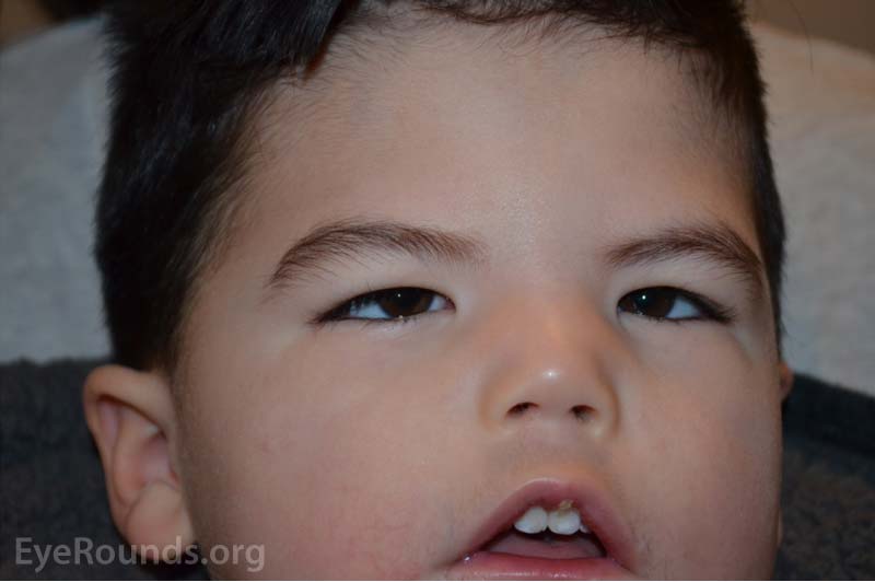
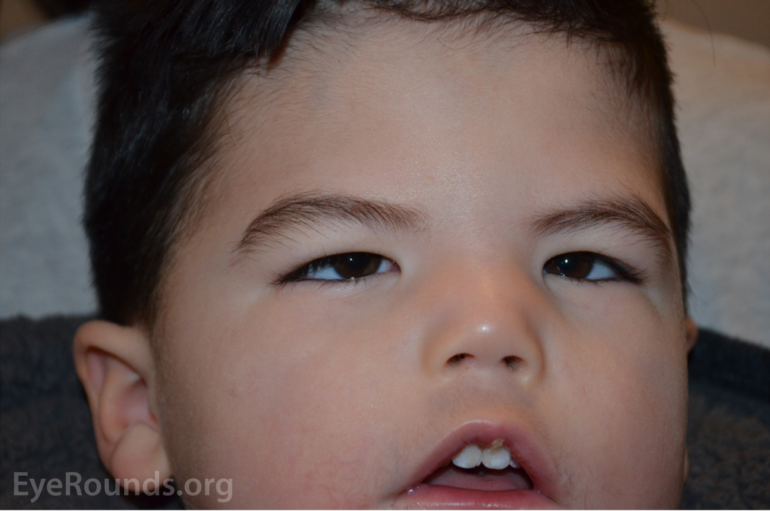
Wide epicanthal folds, decreased facial tone bilaterally, and incomplete mouth closure. Figure 1 demonstrates the described findings as documented in clinical follow up when the child was 2.5 years old.
Differential Diagnosis:
CLINICAL COURSE
The child was followed to monitor for strabismic amblyopia and corneal complications associated with trichiasis. At subsequent follow up, his motility and reduced facial tone remained unchanged. By the age of 2 years, his esotropia stabilized, measuring 45-55 prism diopters. At that point, the patient underwent bilateral medial rectus recessions of 5 mm. While his alignment initially improved, his esotropia recurred and stabilized at 35-45 prism diopters. At this time, the decision was made to inject 5 units of botulinum toxin A into his bilateral medial rectus muscles. This resulted in mild improvement of his alignment, which has remained 20-25 prism diopters at distance and 10 prism diopters at near. Abduction remained limited at -4 in each eye, and adduction was reduced to -2 after his surgery. Over the course of his follow up, his hyperopia has remained within physiological range, and his trichiasis resolved spontaneously. Additionally, given alternate fixation, he was able to demonstrate central, steady, and maintained vision in each eye without the need for patching.
His facial nerve palsy and dysphagia were managed by Otolaryngology. He originally underwent mandibular distraction and cricopharyngeal botulinum toxin A injections to improve his dysphagia and promote oral intake. He gradually improved over the years and transitioned from gastric tube use to oral feeding with fluid intake via his gastric tube.
DIAGNOSIS
Möbius Syndrome
DISCUSSION
Etiology
Möbius syndrome is a disease characterized by congenital palsies of cranial nerves (CN) VI and VII (1), resulting in a non-progressive facial palsy with limitation of ocular abduction. Möbius syndrome was initially described by Von Graefe in 1880 and later studied in more detail by Möbius in 1888 (1). The first Scientific Conference on Möbius Syndrome in 2007 helped establish the "Bethesda Criteria" for diagnosing Möbius Syndrome.
Bethesda Criteria – Möbius Syndrome (2) |
|
Minimum Criteria |
Other Criteria |
|
|
Möbius syndrome is commonly considered a sporadic disease, but several authors have reported cases demonstrating familial inheritance (3-5). Autosomal dominant and autosomal recessive patterns are the most reported.
Carta et al. examined 164 patients with Möbius Syndrome as diagnosed by the Bethesda Criteria and found an estimated prevalence between 0.3-0.6 cases/100,000 live births (6). Within their cohort, the median age of diagnosis was 3.6 years with 44.5% males and 55.5% females (6).
Pathophysiology
The pathogenesis of Möbius syndrome is unclear and remains under debate. One hypothesis postulates that insufficiency of the subclavian artery during the early weeks of gestation, also called the Subclavian Artery Supply Disruption Sequence (SASDS), causes brainstem ischemia due to premature regression or obstruction of the primitive trigeminal arteries before establishment of more mature blood supply from the vertebral arteries
Multiple mechanical factors (thrombus, embolus, connective tissue disorder), embryological events (rapid descent of the heart and great vessels, abnormal configuration of blood vessels, delayed vessel formation), and environmental factors (infection, generalized hypoxia, vasculitis, drugs) have also been postulated as potential etiologies for the pathological changes seen in this syndrome (7).
Further evidence supporting SASDS as a cause has been corroborated in autopsy studies, where investigators observed diffuse brainstem injury in a child with Möbius syndrome who also exhibited paralysis of cranial nerves V, IX, X, XI, and XII. However, SASDS may not fully explain the common extraocular symptoms accompanying Möbius syndrome, as little is known regarding the pathogenesis of clubfeet and hypoplasia of the hands that frequently accompany this syndrome (1).
Signs/Symptoms
CN VI palsy manifests with ocular abduction paralysis, with affected eyes turning inwards. Some reports estimate the presence of bilateral abducens palsies in 91.9% of subjects (8). CN VII palsy manifests as dysfunction of the muscles of facial expression and was noted as bilateral in as much as 82.4% of patients (8). Signs of CN VII palsy include difficulty with eye closure, incomplete mouth closure, and dysarthria, which contribute to the classic masked facies appearance. The following sections describe additional ocular and extraocular anomalies frequently associated with Möbius Syndrome.
Ocular Features
There are three specific patterns of ocular motility alterations: Patterns A (found in 41% of patients), B (50%), and C (9%). Pattern A consists of orthotropia in primary position with complete lack of abduction and adduction (1). Pattern B consists of large angle esotropia, cross fixation, and relative sparing of convergence and adduction (1). Pattern C, the rarest, consists of large angle exotropia in primary position with torticollis, absence of convergence, and vertical eye misalignment (1). Carta et al.'s review of 46 children with Möbius syndrome showed visual acuity is usually good or only moderately impaired. 67% of their subjects possessed binocular function (1). Overall, nearly 46-60% patients have a form of strabismus (mostly esotropia, but occasionally exotropia and hypertropia) (1 ,9).
Extraocular Features
Möbius syndrome can be associated with musculoskeletal pathology such as clubfoot (32%-40%), hypoplastic hands (20%), and hypotonia (75%). Pathology of the oropharyngeal tract results in dysarthria or dysphagia (24-72%), suction defects with feeding difficulties (24%-79%), micrognathia (32%-75%), hearing dysfunction (20%), and microdontia and cleft lip/palate (7-18%) (1 ,8 ,9).
A variety of functional disorders are associated with Möbius Syndrome. The most frequent functional disorders were speech and developmental delays, autism spectrum disorder, and sleep disorders (9 ,10). In addition to dysfunction of CN VI and VII, nearly every other cranial nerve from III–XII has been shown to be impacted by Möbius Syndrome in a mostly equal distribution (21-53%) (9).
Testing/Laboratory Work-Up
Möbius syndrome is exclusively a clinical diagnosis. As with any new patient, a complete eye examination should be performed. Particular attention should be given to ocular movements and visual acuity given the high prevalence of strabismus with possible amblyopia. A complete cranial nerve examination should be completed to identify any additional neurological defects. Additionally, the patient's extremities should be examined to document any anomalies present.
Treatment/Management/Guidelines
Appropriate management of Möbius syndrome requires a multifactorial approach to treat both ocular and extraocular anomalies.
Ocular Anomalies
Patients with Möbius syndrome should continue to follow regularly with Ophthalmology. Strabismus should be managed in a similar manner to other children with strabismus with patching, glasses, and/or surgery as is felt necessary by the pediatric ophthalmologist. Early botulinum toxin injections into the medial rectus have been shown to prevent contractions and help with later strabismus surgery (10). The goal of strabismus surgery in these patients is to improve alignment in primary position, not to restore full ocular motility. These patients should also be evaluated for CN VII palsy-associated exposure keratopathy. If present, treatment involves classic management strategies such as artificial tears, nighttime ointment/gel, surgical intervention, or moisture chambers/goggles/scleral contact lens in more severe disease (11). Finally, sunglasses or hats may be helpful when outside if the child is unable to squint.
Extraocular Anomalies
Management of the extraocular manifestations of Möbius syndrome is multidisciplinary and requires coordination of care with other services.
Summary
|
SIGNS
|
Symptoms
|
Management
|
Bates B, Mortensen Z, Kemp P, De Andrade L. Möebius Syndrome. EyeRounds.org. January 5, 2022. Available from https://EyeRounds.org/cases/319-moebius-syndrome.htmm

Ophthalmic Atlas Images by EyeRounds.org, The University of Iowa are licensed under a Creative Commons Attribution-NonCommercial-NoDerivs 3.0 Unported License.

Address
University of IowaLegal
Related Links
