This site uses tracking information. Visit our privacy policy. Click to agree to this policy and not see this again.

Ophthalmology and Visual Sciences

Familial Exudative Vitreoretinopathy (FEVR)

Familial Exudative Vitreoretinopathy (FEVR)

Joanna I.M. Silverman MD; Mahsaw N. Motlagh MD; Timothy M. Boyce MD; Jonathan F. Russell MD, PhD
August 25, 2022

INITIAL PRESENTATION

Chief Complaint: Bilateral retinal detachments

History of Present Illness:

A 48-day-old boy was referred for evaluation of bilateral retinal detachments. The pregnancy had been complicated by intrauterine growth restriction with a birth weight of 2.6kg. The infant was otherwise healthy. He was first noted to have abnormal red reflexes, non-reactive pupils, and pale discs with concern for overlying vascular abnormalities when evaluated by an outside pediatric ophthalmologist on day 8 of life. An outside retinal surgeon diagnosed bilateral, macula-involving tractional retinal detachments and performed same-day, bilateral lens-sparing vitrectomies. He was seen at the University of Iowa 1 month after surgery. Given his history, an examination under anesthesia (EUA) was performed.

Past Ocular History: Bilateral tractional retinal detachments, status post lens-sparing vitrectomies

Medical History:

  • Born at 37 weeks and 5 days gestation via uncomplicated, spontaneous vaginal delivery
  • Intrauterine growth restriction

Medications: Cholecalciferol

Allergies: No known allergies

Family History: Unremarkable

Social History: Unremarkable

Review of Systems: Unremarkable other than as noted in history of present illness.


OCULAR EXAMINATION

  • Visual Acuity with correction:
    • Right eye (OD): No wince to light
    • Left eye (OS): No wince to light
  • Ocular Motility/Alignment:

    Orthophoric by Hirschberg testing with low amplitude, conjugate nystagmus in the horizontal plane.

  • Intraocular Pressure (IOP):
    • OD: 10 mmHg
    • OS: 9 mmHg
  • Pupils:
    • OD: Non-reactive
    • OS: Non-reactive
  • Confrontation visual fields:
    • OD: Too young to assess
    • OS: Too young to assess
  • B-Scan Echography at presentation (Figure 1):
    • OD: No obvious membranes, localized shallow inferior retinal detachment
    • OS: Hyperechoic membranous elevation inferiorly and temporally consistent with retinal detachment
  • Electroretinography:
    • OD: Non-recordable. Possibly due to young age versus retinal disease.
    • OS: Non-recordable. Possibly due to young age versus retinal disease.
  • External:
    • Normal, both eyes (OU)
  • Slit lamp examination:
    Slit Lamp Exam

    Right Eye

    Left Eye

    Lids/lashes

    Normal

    Normal

    Conjunctiva/sclera

    Normal

    Normal

    Cornea

    Normal

    Normal

    Anterior Chamber

    Deep & quiet

    Deep & quiet

    Iris

    Single posterior synechia at 6 o’clock

    Normal

    Lens

    Clear

    Clear

    Anterior Vitreous

    Normal

    Normal

  • Dilated Fundus Examination (DFE):
    Dilated Fundus Examination (DFE)

    Right Eye

    Left Eye

    Vitreous

    Clear

    Clear

    Disc

    Mild pallor

    Slightly elevated, mild pallor, dragged temporarily

    Cup-to-disc ratio

    0.1

    0.1

    Macula, vessels, and periphery

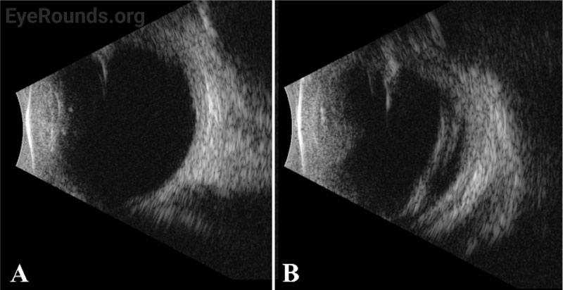
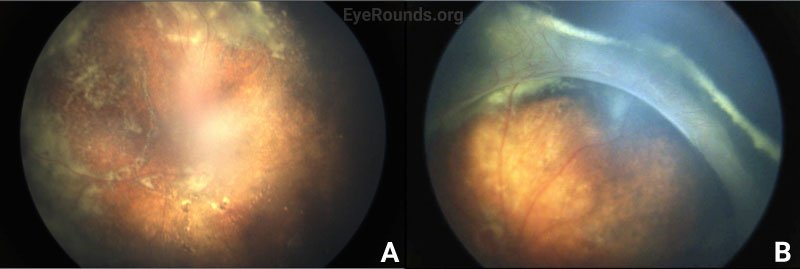
    Vessel dragging/straightening; diffuse pigmentary clumps in the periphery; subretinal exudation with inferior shallow detachment

    Vessel dragging/straightening; diffuse pigmentary clumps in the periphery; less subretinal exudation than right eye concentrated peripherally; 360-degree peripheral anterior loop traction with fibrovascular proliferation, underlying vessels, and traction on peripheral retina

  • B-scan echography, right (A) and left (B) eyes.
    Figure 1. B-scan echography, right (A) and left (B) eyes. The right eye had a localized shallow inferior detachment, while the left eye had a hyperechoic membranous elevation inferiorly and temporally.
    Fundus photographs, right (A) and left (B) eyes.
    Figure 2. Fundus photographs, right (A) and left (B) eyes. Right eye fundus photo demonstrated a distorted macula with diffuse pigmentary clumping and subretinal exudates. Left eye fundus photo showed peripheral anterior loop traction with an opacified vitreous base and fibrovascular proliferation and exudates along this ridge.

    Differential diagnosis:


    DIAGNOSIS: Familial exudative vitreoretinopathy (FEVR)


    CLINICAL COURSE

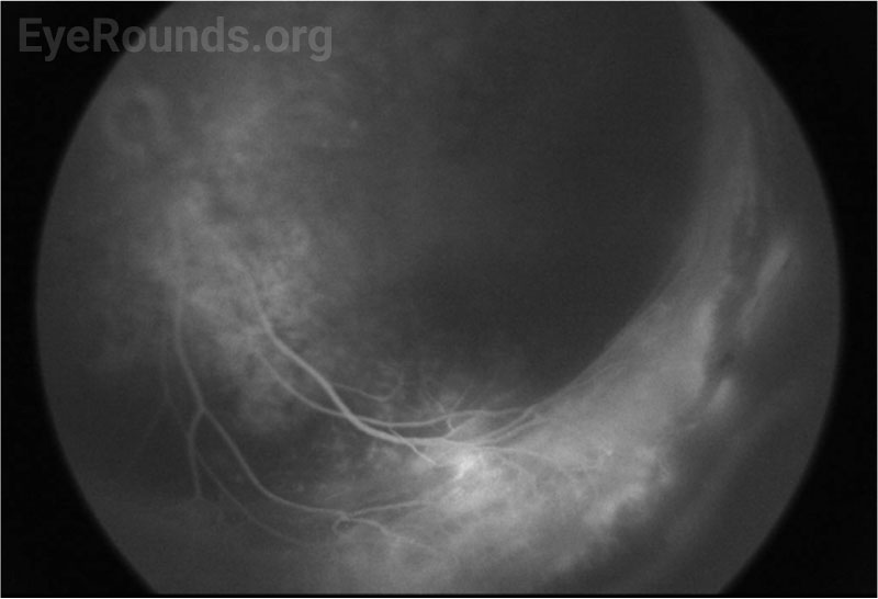
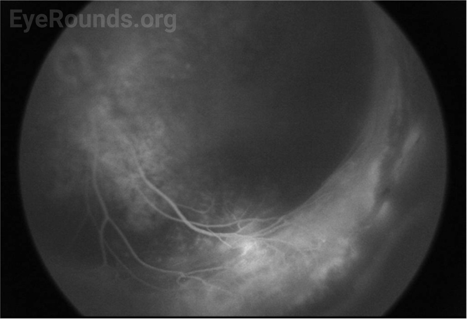
    The patient’s exam findings on EUA were consistent with bilateral familial exudative vitreoretinopathy (FEVR), stage 3B.[1] Operative notes from the outside surgeon indicated that at the time of vitrectomy there was bilateral stage 4B disease. During the EUA, fluorescein angiography demonstrated peripheral neovascularization and ischemia 360 degrees extending to the posterior margin of zone 2 (Figure 3). Given the extensive bilateral neovascularization, panretinal photocoagulation (PRP) was applied to areas of ischemia, which extended from 2–3 disc diameters anterior to the macula up to the visualized fibrovascular ridge. Ablative laser was also applied to areas of shallow retinal detachment that were ischemic but without overlying fibrovascular membranes.

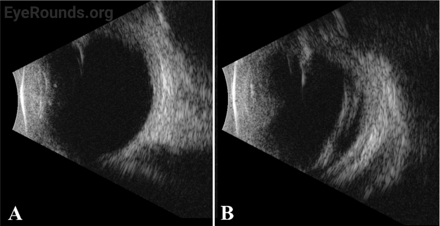
    The patient returned one month later for post-operative evaluation. His mother reported increased responsiveness and tracking to bright light. Fundus exam showed extensive retinal scarring. The retinas were attached except for residual anterior loop traction with peripheral tractional detachment in the left eye. Six months later, the patient was tracking to light, and the retinas remained attached.

    Genetic testing showed an LRP5 mutation (single copy variant of uncertain significance c.685C>T).

    Fluorescein angiography, left eye
    Figure 3. Fluorescein angiography, left eye demonstrating peripheral ischemia and neovascularization 360 degrees extending to posterior zone 2 with a peripheral ridge containing neovascular tufts.

    DISCUSSION

    Etiology/Epidemiology

    FEVR was first reported in 1969 by Criswick and Schepens, who described a progressive, inherited retinal vascular disease resembling retinopathy of prematurity (ROP) that occurred in patients without prematurity.[2]

    Due to its rarity and heterogenous nature, the exact prevalence of FEVR is unknown. There is no gender predilection. Mutations in at least 10 genes (LRP5, FZD4, NDP, ZN408, KIF11, TSPAN12, CTNNB1, JAG1, DOCK6, and ARGHGAP31) have been associated with FEVR.[3-21] However, penetrance and expressivity are variable within families. Only 35-50% of patients with FEVR have a detectable mutation in one of these 10 known causative genes.[1]

    Pathophysiology

    FEVR results from mutations that disrupt signaling in the transduction pathways controlling retinal vascular development, cellular migration, and differentiation. Because of abnormal retinal angiogenesis, there can be exudative retinopathy and/or peripheral retinal ischemia, leading to neovascularization, retinal dragging, and tractional retinal detachment.

    As mentioned above, FEVR is caused by mutations in multiple genes. The inheritance pattern can be autosomal dominant, autosomal recessive, or X-linked, though autosomal dominant is most common.[2,3] Most FEVR genes, including LRP5, FZD4, TSPAN12, and NDP, are involved in signal transduction within the WNT pathway, which regulates retinal vascular development.[2] Additional genes and pathways linked to FEVR are beyond the scope of this review.

    Signs/Symptoms

    FEVR presents with bilateral, abnormal retinal vasculogenesis in children without underlying risk factors for retinopathy of prematurity (i.e., full term, normal birth weight, no supplemental oxygen use). In children, FEVR can be diagnosed because of an abnormal newborn exam, behavioral changes, strabismus/amblyopia, or asymptomatic screening in families carrying a FEVR diagnosis. Between 10-50% of patients have a positive family history.[23] Some affected family members may be asymptomatic but have clear vascular abnormalities on fluorescein angiography.[1]

    FEVR is a clinical diagnosis based on dilated fundus examination, often with supplemental fluorescein angiography. FEVR can feature some or all of the following: avascular peripheral retina (most often the temporal quadrant), dragged retinal vessels and macula (temporally), retinal falciform fold, neovascularization, vitreous hemorrhage, subretinal/intraretinal exudates (occasionally in massive quantities, resembling Coats disease), retinal detachment (which can be exudative, tractional, and/or rhegmatogenous; they occur in 21-64% of patients, and present bilaterally in 68%), persistent fetal vasculature, epiretinal membrane, and peripheral pigmentation (most prominent in the avascular retinal areas).[2,3] As a result of macular temporal dragging, patients may have pseudo-exotropia. Staging criteria for FEVR are shown in Table 1.

    Table 1. Staging criteria for FEVR

    Stage

    Description

    Sub-stage

    A

    B

    1

    Avascular periphery and/or anomalous intraretinal vasculature

    Without exudate

    With exudate

    2

    Avascular periphery with preretinal neovascularization

    Without exudate

    With exudate

    3

    Extramacular retinal detachment

    Without exudate

    With exudate

    4

    Subtotal macula-involving retinal detachment

    Without exudate

    With exudate

    5

    Total retinal detachment

    Open funnel

    Closed funnel

    Not all cases of FEVR are symptomatic or progressive.[4] Some patients are not diagnosed with FEVR until adolescence, when the disease often progresses. Rarely, a patient is not diagnosed until adulthood, such as if they are screened because of a FEVR diagnosis in a symptomatic relative. The average age at diagnosis is 58.6 months.[3] Severity is often inversely proportional to age of diagnosis. FEVR rarely progresses in adulthood, although vitreous hemorrhage and other vision-threatening complications can occur.[5] Thus, long term follow-up is imperative.

    Testing/Laboratory work-up

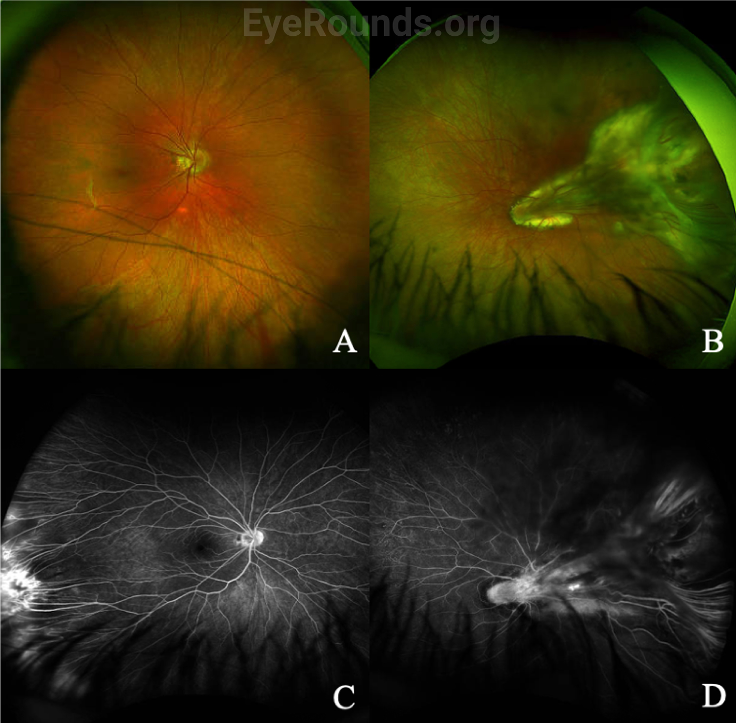
    In addition to a thorough dilated fundus exam, which may require EUA in children, wide-angle fluorescein angiography (FA) is helpful in diagnosing FEVR, especially in milder cases where only the far peripheral retina is affected. Typical findings on fluorescein angiography include a V-shaped avascular zone in the temporal periphery.[1] Peripheral avascularity can extend 360 degrees in more severe cases. Additional evidence on FA that suggests FEVR includes vessel straightening, peripheral telangiectasias, neovascularization, and small-vessel fluorescein leakage, as shown for a separate patient in Figure 4.[2]

    Fundus photography and fluorescein angiography of FEVR, both eyes
    Figure 4. Fundus photography and fluorescein angiography of FEVR in an 18-year-old, right (A and C) and left (B and D) eyes. Although the fundus photo of the right eye shows only mild temporal vessel straightening, the FA shows peripheral non-perfusion, preretinal neovascularization, and vessel leakage. The left eye has a prominent falciform fold coursing from the optic nerve through the fovea to the temporal periphery, subretinal exudates, and vitreous bands overlying a tractional retinal detachment in the temporal periphery. The FA shows vessel straightening, peripheral nonperfusion with late, largely small-vessel leakage, staining in the area of exudation, and nasal PRP scars. This patient has stage 2A disease in the right eye and stage 4B disease in the left eye.

    Genetic testing is important to verify the diagnosis and to counsel patients and families. However, since fewer than 50% of FEVR cases can be attributed to a known gene, a negative genetic test result should not rule out a diagnosis of FEVR.[2]

    Treatment/Management

    The management of FEVR depends on the stage of disease. Stage 1 disease can be observed, while Stage 2 FEVR is usually treated with laser photocoagulation of avascular zones to decrease risk of exudation and neovascular complications such as vitreous hemorrhage and retinal detachment.[2] Compared to anti-VEGF agents, laser remains the mainstay of treatment, though anti-VEGF injections are sometimes used adjunctively, especially in the presurgical period to minimize intraoperative hemorrhage.[2,3] Treatment for stage 3-5 FEVR depends on the cause of retinal detachment. If exudative in nature, laser to avascular retina can be effective. Tractional and rhegmatogenous retinal detachments usually require retinal surgery; in infants, this is usually lens-sparing vitrectomy, whereas older children can often be treated with scleral buckling.

    Genetic testing results inform screening of asymptomatic family members, a small percentage of whom may require treatment. Counseling female carriers of child-bearing age can facilitate pre-implantation genetic diagnosis and/or prompt screening of their newborns.

    EPIDEMIOLOGY AND ETIOLOGY

    • Abnormal retinal angiogenesis during early development
    • Prevalence unknown
    • 35-50% of patients carry mutations in LRP5, FZD4, NDP, ZN408, KIF11, TSPAN12, CTNNB1, JAG1, DOCK6, ARGHGAP31, ATOH7, or RCGTB1

    DIAGNOSIS

    • DFE/EUA
    • Fluorescein angiography
    • Genetic testing

    SIGNS/SYMPTOMS

    • Avascular peripheral retina in a full-term, normal weight infant without supplemental oxygen use
    • Additional signs include retinal vessel straightening, macular dragging, subretinal exudates, preretinal neovascularization, retinal detachment, and pseudo-exotropia
    • Preverbal status of many patients leads to diagnosis after abnormal newborn exam, change in behavior, or known family history

    TREATMENT/MANAGEMENT

    • Stage 1A and 1B: observation
    • Stage 2: laser photocoagulation, and/or anti-VEGF
    • Stage 3-5: laser, anti-VEGF, and/or retinal surgery

    References

    • 1. Kashani AH, Learned D, Nudleman E, Drenser KA, Capone A, Trese MT. High prevalence of peripheral retinal vascular anomalies in family members of patients with familial exudative vitreoretinopathy. Ophthalmology 2014;121(1):262-268. [PMID 24084499]
    • 2. Criswick VG, Schepens CL. Familial exudative vitreoretinopathy. Am J Ophthalmol 1969;68(4):578-594. [PMID 5394449]
    • 3. Robitaille J, MacDonald ML, Kaykas A, Sheldahl LC, Zeisler J, Dubé MP, Zhang LH, Singaraja RR, Guernsey DL, Zheng B, Siebert LF, Hoskin-Mott A, Trese MT, Pimstone SN, Shastry BS, Moon RT, Hayden MR, Goldberg YP, Samuels ME. Mutant frizzled-4 disrupts retinal angiogenesis in familial exudative vitreoretinopathy. Nat Genet 2002;32(2):326-330. [PMID 12172548]
    • 4. Toomes C, Bottomley HM, Jackson RM, Towns KV, Scott S, Mackey DA, Craig JE, Jiang L, Yang Z, Trembath R, Woodruff G, Gregory-Evans CY, Gregory-Evans K, Parker MJ, Black GC, Downey LM, Zhang K, Inglehearn CF. Mutations in LRP5 or FZD4 underlie the common familial exudative vitreoretinopathy locus on chromosome 11q. Am J Hum Genet 2004;74(4):721-730. [PMID 15024691]
    • 5. Plager DA, Orgel IK, Ellis FD, Hartzer M, Trese MT, Shastry BS. X-linked recessive familial exudative vitreoretinopathy. Am J Ophthalmol 1992;114(2):145-148. [PMID 1642288]
    • 6. Chen ZY, Battinelli EM, Fielder A, Bundey S, Sims K, Breakefield XO, Craig IW. A mutation in the Norrie disease gene (NDP) associated with X-linked familial exudative vitreoretinopathy. Nat Genet 1993;5(2):180-183. [PMID 8252044]
    • 7. Gong Y, Slee RB, Fukai N, Rawadi G, Roman-Roman S, Reginato AM, Wang H, Cundy T, Glorieux FH, Lev D, Zacharin M, Oexle K, Marcelino J, Suwairi W, Heeger S, Sabatakos G, Apte S, Adkins WN, Allgrove J, Arslan-Kirchner M, Batch JA, Beighton P, Black GC, Boles RG, Boon LM, Borrone C, Brunner HG, Carle GF, Dallapiccola B, De Paepe A, Floege B, Halfhide ML, Hall B, Hennekam RC, Hirose T, Jans A, Jüppner H, Kim CA, Keppler-Noreuil K, Kohlschuetter A, LaCombe D, Lambert M, Lemyre E, Letteboer T, Peltonen L, Ramesar RS, Romanengo M, Somer H, Steichen-Gersdorf E, Steinmann B, Sullivan B, Superti-Furga A, Swoboda W, van den Boogaard MJ, Van Hul W, Vikkula M, Votruba M, Zabel B, Garcia T, Baron R, Olsen BR, Warman ML. LDL receptor-related protein 5 (LRP5) affects bone accrual and eye development. Cell 2001;107(4):513-523. [PMID 11719191]
    • 8. Jiao X, Ventruto V, Trese MT, Shastry BS, Hejtmancik JF. Autosomal recessive familial exudative vitreoretinopathy is associated with mutations in LRP5. Am J Hum Genet 2004;75(5):878-884. [PMID 15346351]
    • 9. Poulter JA, Ali M, Gilmour DF, Rice A, Kondo H, Hayashi K, Mackey DA, Kearns LS, Ruddle JB, Craig JE, Pierce EA, Downey LM, Mohamed MD, Markham AF, Inglehearn CF, Toomes C. Mutations in TSPAN12 Cause Autosomal-Dominant Familial Exudative Vitreoretinopathy. Am J Hum Genet 2016;98(3):592. [PMID 28863275]
    • 10. Nikopoulos K, Gilissen C, Hoischen A, van Nouhuys CE, Boonstra FN, Blokland EA, Arts P, Wieskamp N, Strom TM, Ayuso C, Tilanus MA, Bouwhuis S, Mukhopadhyay A, Scheffer H, Hoefsloot LH, Veltman JA, Cremers FP, Collin RW. Next-generation sequencing of a 40 Mb linkage interval reveals TSPAN12 mutations in patients with familial exudative vitreoretinopathy. Am J Hum Genet 2010;86(2):240-247. [PMID 20159111]
    • 11. Poulter JA, Davidson AE, Ali M, Gilmour DF, Parry DA, Mintz-Hittner HA, Carr IM, Bottomley HM, Long VW, Downey LM, Sergouniotis PI, Wright GA, MacLaren RE, Moore AT, Webster AR, Inglehearn CF, Toomes C. Recessive mutations in TSPAN12 cause retinal dysplasia and severe familial exudative vitreoretinopathy (FEVR). Invest Ophthalmol Vis Sci 2012;53(6):2873-2879. [PMID 22427576]
    • 12. Collin RW, Nikopoulos K, Dona M, Gilissen C, Hoischen A, Boonstra FN, Poulter JA, Kondo H, Berger W, Toomes C, Tahira T, Mohn LR, Blokland EA, Hetterschijt L, Ali M, Groothuismink JM, Duijkers L, Inglehearn CF, Sollfrank L, Strom TM, Uchio E, van Nouhuys CE, Kremer H, Veltman JA, van Wijk E, Cremers FP. ZNF408 is mutated in familial exudative vitreoretinopathy and is crucial for the development of zebrafish retinal vasculature. Proc Natl Acad Sci U S A 2013;110(24):9856-9861. [PMID 23716654]
    • 13. Robitaille JM, Gillett RM, LeBlanc MA, Gaston D, Nightingale M, Mackley MP, Parkash S, Hathaway J, Thomas A, Ells A, Traboulsi EI, Héon E, Roy M, Shalev S, Fernandez CV, MacGillivray C, Wallace K, Fahiminiya S, Majewski J, McMaster CR, Bedard K. Phenotypic overlap between familial exudative vitreoretinopathy and microcephaly, lymphedema, and chorioretinal dysplasia caused by KIF11 mutations. JAMA Ophthalmol 2014;132(12):1393-1399. [PMID 25124931]
    • 14. Hu H, Xiao X, Li S, Jia X, Guo X, Zhang Q. KIF11 mutations are a common cause of autosomal dominant familial exudative vitreoretinopathy. Br J Ophthalmol 2016;100(2):278-283. [PMID 26472404]
    • 15. Chen C, Sun L, Li S, Huang L, Zhang T, Wang Z, Yu B, Luo X, Ding X. Novel variants in familial exudative vitreoretinopathy patients with KIF11 mutations and the Genotype-Phenotype correlation. Exp Eye Res 2020;199:108165. [PMID 32730767]
    • 16. Li JK, Fei P, Li Y, Huang QJ, Zhang Q, Zhang X, Rao YQ, Li J, Zhao P. Identification of novel KIF11 mutations in patients with familial exudative vitreoretinopathy and a phenotypic analysis. Sci Rep 2016;6:26564. [PMID 27212378]
    • 17. Sun W, Xiao X, Li S, Jia X, Wang P, Zhang Q. Germline Mutations in CTNNB1 Associated With Syndromic FEVR or Norrie Disease. Invest Ophthalmol Vis Sci 2019;60(1):93-97. [PMID 30640974]
    • 18. Dixon MW, Stem MS, Schuette JL, Keegan CE, Besirli CG. CTNNB1 mutation associated with familial exudative vitreoretinopathy (FEVR) phenotype. Ophthalmic Genet 2016;37(4):468-470. [PMID 26967979]
    • 19. Zhang L, Zhang X, Xu H, Huang L, Zhang S, Liu W, Yang Y, Fei P, Li S, Yang M, Zhao P, Zhu X, Yang Z. Exome sequencing revealed Notch ligand JAG1 as a novel candidate gene for familial exudative vitreoretinopathy. Genet Med 2020;22(1):77-84. [PMID 31273345]
    • 20. Wu JH, Liu JH, Ko YC, Wang CT, Chung YC, Chu KC, Liu TT, Chao HM, Jiang YJ, Chen SJ, Chung MY. Haploinsufficiency of RCBTB1 is associated with Coats disease and familial exudative vitreoretinopathy. Hum Mol Genet 2016;25(8):1637-1647. [PMID 26908610]
    • 21. Kondo H, Matsushita I, Tahira T, Uchio E, Kusaka S. Mutations in ATOH7 gene in patients with nonsyndromic congenital retinal nonattachment and familial exudative vitreoretinopathy. Ophthalmic Genet 2016;37(4):462-464. [PMID 26933893]
    • 22. Robitaille JM, Zheng B, Wallace K, Beis MJ, Tatlidil C, Yang J, Sheidow TG, Siebert L, Levin AV, Lam WC, Arthur BW, Lyons CJ, Jaakkola E, Tsilou E, Williams CA, Weaver RG, Jr., Shields CL, Guernsey DL. The role of Frizzled-4 mutations in familial exudative vitreoretinopathy and Coats disease. Br J Ophthalmol 2011;95(4):574-579. [PMID 21097938]
    • 23. Gilmour DF. Familial exudative vitreoretinopathy and related retinopathies. Eye (Lond) 2015;29(1):1-14. [PMID 25323851]
    • 24. Clevers H. Eyeing up new Wnt pathway players. Cell 2009;139(2):227-229. [PMID 19837026]
    • 25. Ranchod TM, Ho LY, Drenser KA, Capone A, Jr., Trese MT. Clinical presentation of familial exudative vitreoretinopathy. Ophthalmology 2011;118(10):2070-2075. [PMID 21868098]
    • 26. Ober RR, Bird AC, Hamilton AM, Sehmi K. Autosomal dominant exudative vitreoretinopathy. Br J Ophthalmol 1980;64(2):112-120. [PMID 7362811]
    • 27. Tagami M, Kusuhara S, Honda S, Tsukahara Y, Negi A. Rapid regression of retinal hemorrhage and neovascularization in a case of familial exudative vitreoretinopathy treated with intravitreal bevacizumab. Graefes Arch Clin Exp Ophthalmol 2008;246(12):1787-1789. [PMID 18795314]
    • 28. Miyakubo H, Hashimoto K, Miyakubo S. Retinal vascular pattern in familial exudative vitreoretinopathy. Ophthalmology 1984;91(12):1524-1530. [PMID 6084219]
    • 29. Salvo J, Lyubasyuk V, Xu M, Wang H, Wang F, Nguyen D, Wang K, Luo H, Wen C, Shi C, Lin D, Zhang K, Chen R. Next-generation sequencing and novel variant determination in a cohort of 92 familial exudative vitreoretinopathy patients. Invest Ophthalmol Vis Sci 2015;56(3):1937-1946. [PMID 25711638]
    • 30. Pendergast SD, Trese MT. Familial exudative vitreoretinopathy. Results of surgical management. Ophthalmology 1998;105(6):1015-1023. [PMID 9627651]
    • 31. Henry CR, Sisk RA, Tzu JH, Albini TA, Davis JL, Murray TG, Berrocal AM. Long-term follow-up of intravitreal bevacizumab for the treatment of pediatric retinal and choroidal diseases. J aapos 2015;19(6):541-548. [PMID 26691034]

Suggested citation format:

Silverman JIM, Motlagh MN, Boyce TM, Russell JF. Familial Exudative Vitreoretinopathy (FEVR). EyeRounds.org. August 25, 2022. Available from https://eyerounds.org/cases/333-FEVR.htm


Image Permissions:
Creative Commons

Ophthalmic Atlas Images by EyeRounds.org, The University of Iowa are licensed under a Creative Commons Attribution-NonCommercial-NoDerivs 3.0 Unported License.