Chief Complaint: 29-year-old male with HA, hematuria, and visual obscurations.
History of Present Illness: 29-year-old male complained of headache (HA) for several weeks. HA was worse the day before presentation (8/10) and was associated with nausea & vomiting (N/V). He went to the local ER and was noted to have hematuria. The patient was told he was "dehydrated" from the N/V, given pain meds, and sent home.
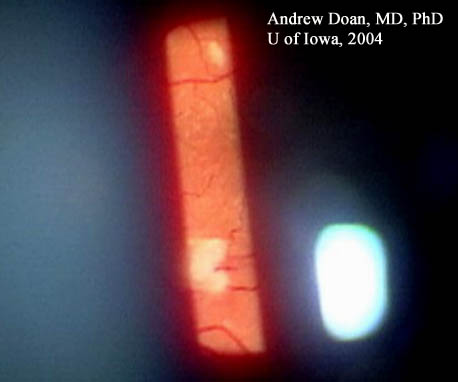
On the next day, he awoke feeling weak and complained of HA and N/V. He noticed visual field (VF) obscurations in the left eye (OS). He then presented for formal ophthalmology evaluation.
PMH: Poorly controlled HTN (160/108) with losartan & amlodipine.
EXAM:
Urinalysis indicated 3+ hematuria and proteinuria. Not gross. CBC: |
ESR 1 mm/hr HIV NEGATIVE Head CT negative |
His electrolyte panel was notable for: |
The peripheral blood smear showed: LDH was 752. |
The patient was admitted, and the following day, his hemoglobin dropped from 14.1 g/dl to 9 g/dl.
Although a nonspecific finding, the presence of cotton wool spots in an ill, young patient should elicit a careful review of systems and consideration of a thorough medical evaluation. Table 1 details common and uncommon diagnoses for cotton wool spots of the retina.
Given the clinical presentation, our patient was diagnosed with thrombotic thrombocytopenic purpura-hemolytic uremic syndrome (TTP-HUS), see Table 2. Although, once thought to be separate pathologic entities, it is now felt that these entities represent a spectrum of a single disorder. TTP-HUS presents as a thrombocytopenia and microangiopathic hemolytic anemia associated with thrombi composed primarily of platelets in affected organs. Patients may present with: thrombocytopenia, microangiopathic hemolytic anemia, neurologic signs and symptoms (e.g., headache, vision changes, paresthesias), renal insufficiency, and fever. TTP-HUS is a deadly disorder with mortality rates over 90% in untreated patients. Plasma exchange with fresh frozen plasma is the treatment of choice and reduces the mortality rate to below 50%. Patient survival is dependent on prompt diagnosis, hospital admission, and treatment.
While this patient came to our attention due to a new scotoma in his visual field from large cotton wool spots, other ophthalmic manifestations in patients with TTP-HUS include: serous macular detachments, choriocapillaris occlusion due to fibrin-platelet thrombi, retinal or vitreous hemorrhages, optic disc edema, neovascularization of the disc, and central retinal vein occlusion.
Cotton wool spots can be a sign of serious systemic disease and warrant further evaluation in certain clinical scenarios. Initial testing should include blood pressure measurement, complete blood count with platelets and differential, and evaluation for diabetes. In this patient, a life-threatening illness was discovered. Although TTP-HUS is a rare disorder, it should be considered in patients who present with vaso-occlusive disease of the retina, neurologic decline, renal insufficiency, and hematologic abnormalities (such as anemia and/or thrombocytopenia). Evaluation and management of TTP-HUS should be done in conjunction with internists and hematologists. Because the mortality rate of this condition can be exceedingly high without prompt intervention, ophthalmologists may play a key role in its timely diagnosis and thus improve the likelihood of patient survival.
COMMON
|
LESS COMMON
|
Symptoms
|
Treatment
|
Age and Sex
|
Long-Term Outcomes
|
Suggested citation format: Doan A, Farjo A: TTP-HUS: 29-year-old male with HA, hematuria, and visual obscurations. February 21, 2005; Available from: http://www.EyeRounds.org/cases/case1.htm.

Ophthalmic Atlas Images by EyeRounds.org, The University of Iowa are licensed under a Creative Commons Attribution-NonCommercial-NoDerivs 3.0 Unported License.

Address
University of IowaLegal
Related Links