This site uses tracking information. Visit our privacy policy. Click to agree to this policy and not see this again.

Ophthalmology and Visual Sciences

Verticillata:

54-year-old white male with a known history of atrial fibrillation and hypertension on amiodarone

Verticillata:

54-year-old white male with a known history of atrial fibrillation and hypertension on amiodarone
Jordan M. Graff, MD
February 21, 2005

Chief Complaint: Patient on Amiodarone therapy. No visual complaints.

History of Present Illness: 54-year-old white male with a known history of atrial fibrillation and hypertension. The patient had been on Amiodarone therapy for several years. Two months prior to this routine follow up, the patient's primary care doctor made a change in the patient's cardiac medications which included the removal of amiodarone from his medication regimen. The patient has no vision complaints.

PMH/POH: Atrial fibrillation and hypertension. No other ocular or health problems.

EXAM, OCULAR:

  • VA 20/20 OD and OS
  • Intraocular Pressures and Confrontation Visual Fields normal
  • DFE normal OU
  • EOM Full OU
  • SLE notable for corneal deposits in the cornea at the level of the basal epithelium, inferiorly OU. Deposits form a faint golden-brown whorl pattern evident in both corneas (see Figure 1).
Figure 1: Whorled pattern of corneal verticillata on the inferior cornea in patient with chronic amiodarone use.

Figure 2: Another example of similar findings in a patient taking amiodarone.

Discussion

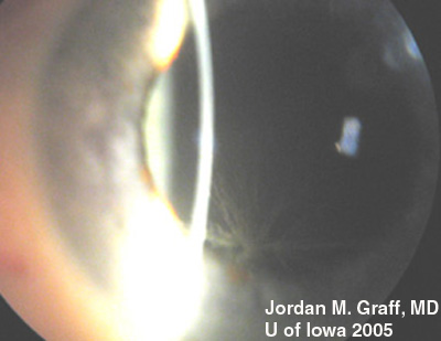
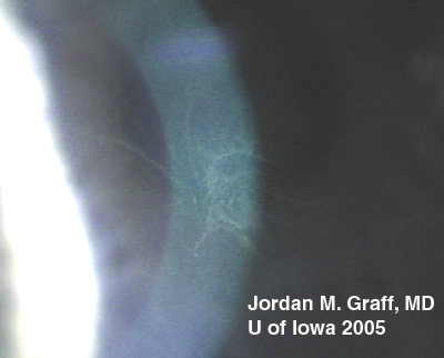
There are dozens of medications, materials, and disease biproducts that can result in deposits in the cornea. These deposits occur in various levels of the cornea. Corneal verticillata, also called vortex keratopathy, are one type of corneal deposits. Verticillata are readily recognizable by their distinct appearance as fine, golden-brown or gray opacities that are almost always bilateral. The opacities branch out from a central whorl, usually across the inferior cornea. The most common causes of verticillata include:

  • Amiodarone
  • Chloroquine and Hydroxychloroquine
  • Indomethacin
  • Phenothiazines
  • Fabry disease (see Figure 3)
Figure 3: Fabry disease with corneal verticillata

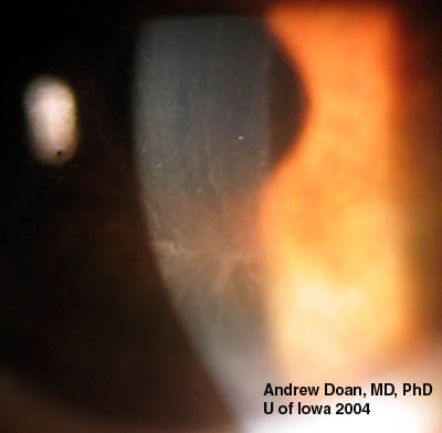
In the case of our patient, he had developed classic corneal verticillata from amiodarone use. As can be seen, the verticillata are somewhat less distinct than some published examples. Having been off the medication for 2 months, it is expected that the deposition pattern will diminish, as is the case for the vast majority of patients. Verticillata almost never cause vision complaints. Rarely, patients may notice haloes. The deposits occur within the subepithelium, do not stain, and are non-irritating. It has been hypothesized that the amphiphilic drugs that deposit verticillata may form complexes with phospholipids in the cells. These complexes cannot be metabolized by lysosomal phospholipases. Tissues where phospholipids content or turnover is high, lamellar or crystalloid deposits may occur. Verticillata from indomethacin, amiodarone, chloroquines, and phenothiazines are similar in this respect.

Verticillata from Fabry disease are similar in appearance to those caused by medication related deposits. Fabry disease is a glycolipidosis caused by deficiency of the alpha-galactosidase A (alpha-Gal A) enzyme. In addition to the verticillata and corneal spoke-like deposits, patients often have severe pain in the extremities, and characteristic skin lesions (angiokeratomas). In the third or fourth decade of life, more severe effects of the disease are manifest including progressive cardiovascular, renal, and cerebrovascular disease (seizures and strokes). Some heterozygote female carriers of the alpha-Gal A defect may express varying degrees of the disease as carriers, including corneal verticillata.

Diagnosis: Corneal Verticillata

EPIDEMIOLOGY

  • There is no data to suggest protective nor inducing factors in patients who develop verticillata from medication use.
  • Most studies suggest that all or nearly all patients taking amiodarone will develop verticillata.
  • Regarding Fabry disease as a separate entity, it is a rare disorder (1:40,000) with no race predilection.

SIGNS

  • Fine, golden-brown or gray opacities branch out from a central whorl, usually across the inferior cornea.
  • Almost always bilateral
  • Deposits occur in the basal epithelium

SYMPTOMS

  • Patients generally have no symptoms from verticillata
  • Few patients may notice haloes

TREATMENT

  • There is no recommended treatment for verticillata and medication regimens are not altered based on the presence of corneal deposits.
  • When depositing drugs are stopped for other reasons, as in the case here presented, most verticillata will eventually fade away.

Differential Diagnoses for corneal verticillata:

  • Iron lines (ie. after radial keratotomy)
  • Epithelial defect
  • Stromal deposits (Gold, Silver, Antacid, Retinoid depositions)
  • Amiodarone
  • Chloroquine and Hydroxychloroquine
  • Indomethacin
  • Phenothiazines
  • Fabry disease
References
  1. Chew E, Ghosh M, McCulloch C. Amiodarone-induced cornea verticillata. Can J Ophthalmol. 1982;17(3):96-9.
  2. Hollander DA, Aldave AJ. Drug-induced corneal complications. Curr Opin Ophthalmol. 2004;15(6):541-8.
  3. Arffa RC, Eve FR. Systemic associations of corneal deposits. Int Ophthalmol Clin. 1991;31(3):89-110.
  4. Masson C, Cisse I, Simon V, Insalaco P, Audran M. Fabry disease: a review. Joint Bone Spine. 2004;71(5):381-3.
  5. Orssaud C, Dufier J, Germain D. Ocular manifestations in Fabry disease: a survey of 32 hemizygous male patients. Ophthalmic Genet. 2003;24(3):129-39.
  6. Clinical Approach to Depositions and Degenerations of the Conjunctiva, Cornea, and Sclera. In Sutphin Jr. JE, Chodosh H, Dana MR, Fowler WC, Reidy JJ, Weiss JS, Turgeon PW, External Disease and Cornea. Section 8, Basic and Clinical Science Course. San Francisco: American Academy of Ophthalmology; 2004; Chapter 17, p. 353.
Suggested citation format:

Suggested citation format: Graff JM. Verticillata: 54-year-old white male with a known history of atrial fibrillation and hypertension on amiodarone. EyeRounds.org, February 21, 2005; Available from http://www.EyeRounds.org/cases/case29.htm


last updated: 02-21-2005

Image Permissions:
Creative Commons

Ophthalmic Atlas Images by EyeRounds.org, The University of Iowa are licensed under a Creative Commons Attribution-NonCommercial-NoDerivs 3.0 Unported License.