This site uses tracking information. Visit our privacy policy. Click to agree to this policy and not see this again.

Ophthalmology and Visual Sciences

Basal Cell Carcinoma, Morpheaform subtype:

78-year-old male with complaints of enlarging Left Lower Lid lesion

Basal Cell Carcinoma, Morpheaform subtype:

78-year-old male with complaints of enlarging Left Lower Lid lesion
Andrew Doan, MD, PhD, Thomas Oetting, MD
February 21, 2005

Chief Complaint:. 78-year-old male with complaints of enlarging LLL lesion.

History of Present Illness: Patient complained of a painless, red lesion noticed on the left lower lid (LLL) for 4-6 months. He was not sure if the lesion had grown in size, but wanted an evaluation because the lesion has not resolved.

PMH: Hypertension, mild coronary artery disease, and mild cataracts.

Exam:
  • Best corrected visual acuities: 20/25 OU.
  • Pupils: equal, no RAPD.
  • EOM: full OU
  • VF: normal
  • DFE: retina exam notable for normal macula, vessels, and periphery OU.
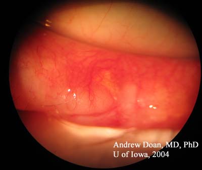
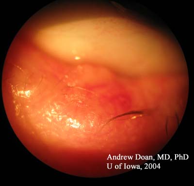
  • SLE: notable for the lesion shown below LLL. Mild cataracts OU.
LLL Lesion
Photo of LLL lesion
SLE LLL
SLE Photo of the LLL lesion (higher magnification). Note the loss of eye lashes (madarosis) and irregular lid margin.
SLE LLL
SLE Photo of the LLL lesion, lid margin & palpebral conjunctivae. Note the telangiectasia of the lesion, disruption of the lid margin, and hyperemia. Borders of this lesion are indistinct.

Discussion

Morpheaform Basal Cell Carcinoma

This patient was diagnosed initially with a chalazion. When warm compresses and lid hygiene failed to resolve the LLL lesion, a full-thickness wedge biopsy was performed. We should be concerned about sebaceous cell carcinoma (SCC) and masquerade syndrome because SCC can be difficult to diagnose and detect. If one is concerned about SCC, the ocular pathologist should be informed so that oil red-O stain will be used to detect presence of lipid. Because SCC can be missed on frozen section, half of the specimen was sent for frozen section evaluation and the other half of the specimen was sent for histopathology.

The pathology report was consistent with morpheaform basal cell carcinoma (BCC). In morpheaform BCC, instead of bulky tumor nodules, there are slender strands of tumor cells. Morpheaform BCC is the most aggressive of all the BCC types: nodulo-ulcerative (most common), pigmented, morpheaform, and superficial.


Diagnosis: Morpheaform Basal Cell Carcinoma

EPIDEMIOLOGY

  • Incidence: 300-600 cases per 100,000
  • 30% lifetime risk
  • Age: adults and elderly
  • Male:Female ratio 3:2
  • Low mortality, but high morbidity due to disfigurement.
  • Most common skin cancer.

SIGNS

  • Nodulo-ulcerative: waxy papules with central depression, ulceration, bleeding, crusting, rolled border, translucency, and telangiectases over lesion.
  • Pigmented: areas of brown and black pigment seen in lesion along with the signs similar to nodulo-ulcerative.
  • Morpheaform: sclerotic plaques or papules, border not defined, and bleeding/ucleration/crusting are not common.
  • Superficial: scaly patches or papules, threadlike border, and can mimic psoriasis or eczema.
  • Madarosis (loss of lashes)
  • Disruption of lid margin and architecture

SYMPTOMS

  • Non-healing sore which may bleed of varying duration on face, ears, lids, neck, and skin.
  • Bleeding with minor trauma
  • Ulceration
  • Sun exposure
  • Radiation exposure
  • Arsenic exposure
  • h/o Xeroderma pigmentosum

TREATMENT

  • Take wedge biopsy to rule-out sebaceous cell carcinoma.
  • Mohs' micrographic surgery or excision with frozen-section control.
  • Close follow-up for recurrence.

Differential Diagnoses:

  • Actinic Keratosis
  • Bowen Disease
  • Keratoacanthoma
  • Sebaceous Hyperplasia
  • Seborrheic Keratosis
  • Squamous Cell Carcinoma
  • Trichoepithelioma

References
  1. Lear JT, Smith AG. Basal cell carcinoma. Postgrad Med J. 1997 Sep;73(863):538-42.
  2. Michael L Ramsey, MD. Basal Cell Carcinoma. www.emedicine.com
  3. Nerad JA, Whitaker DC. Am J Ophthalmol. 1988 Dec 15;106(6):723-9. Periocular basal cell carcinoma in adults 35 years of age and younger.
Suggested citation format:

Doan A, Oetting TA:Basal Cell Carcinoma, Morpheaform subtype: 78-year-old male with complaints of enlarging left lower lid lesion. February 21, 2005; Available from: http://www.EyeRounds.org/cases/case7.htm.


last updated: 02-21-2005

Image Permissions:
Creative Commons

Ophthalmic Atlas Images by EyeRounds.org, The University of Iowa are licensed under a Creative Commons Attribution-NonCommercial-NoDerivs 3.0 Unported License.