

Digital Photography
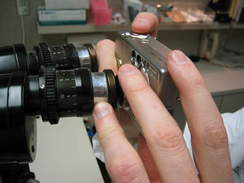
Some of the photos were obtained with a Canon Powershot Digital Elph (we recommend the models with image stabilization) compact digital camera through the ocular of the slit lamp biomicroscope or operating microscope. The fundus photographs were obtained from a digital photography system made by OIS.
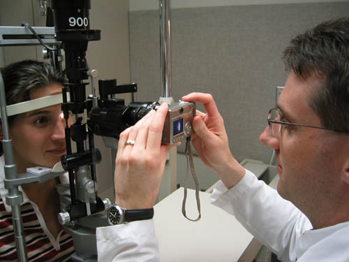
Dr. Michael Boland taking a picture of Dr. Lynn Fraterrigo's eye.
To take detailed images through the slit lamp ocular:
-
Use a secondary diffuse light (optional).
-
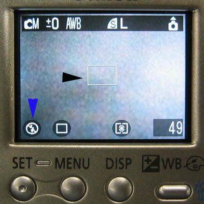
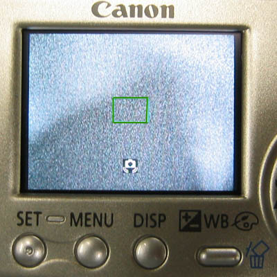
Set the camera to center focus mode (Black Arrow). Turn off flash photography (Blue Arrow).
-
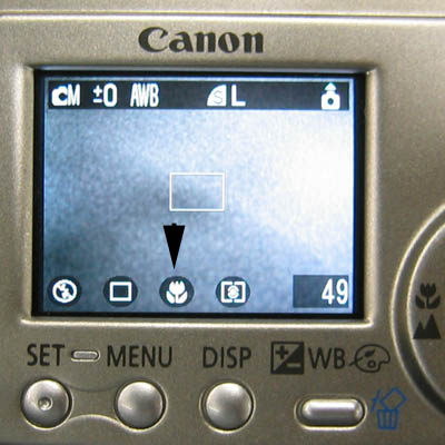
Set the camera to MACRO function.
-
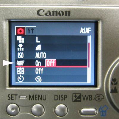
Turn off AiAF
-
Position the camera in front of the ocular until the camera is able to focus on the image (Center Box Turns Green while the shutter button is pressed halfway). Press the shutter button all the way down to capture image.
-
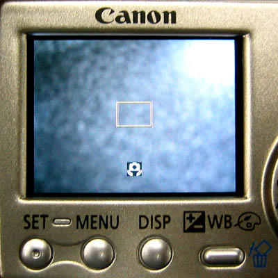
If Center Box Turns Yellow while the shutter button is pressed halfway, then reposition camera and refocus again with the shutter button pressed halfway.
-
With practice, high resolution images can be obtained of the anterior segment.
More about digital photography in ophthalmology