Age-related macular degeneration (AMD) is the leading cause of blindness in elderly patients [1] and is classified as either non-exudative (i.e., dry) or exudative (i.e., wet or neovascular). The clinical manifestations of non-exudative AMD range from incidental findings of drusen to geographic atrophy causing significant vision loss, and approximately 15% of AMD patients ultimately transition to the wet form [2]. This conversion is due to the extension of choroidal vessels into the sub-retinal pigment epithelial (sub-RPE) or sub-retinal space via breaks in Bruch's membrane. AMD has been categorized by The Age-Related Eye Disease Study (AREDS) based on exam findings of hard drusen, soft drusen, RPE abnormalities, atrophy, and choroidal neovascularization [3]; the AREDS categories are as follows:
Category 1 (No AMD): a few (5-15), small (<63µm) or no drusen without pigment changes.
Category 2 (Early AMD): Several small drusen, few intermediate-sized (63-124µm) drusen, and/or pigmentary changes in one or both eyes
Category 3 (Intermediate AMD): Extensive (i.e., 20 soft or 65 hard without any soft) intermediate-sized drusen, one large (>125µm) druse, and/or geographic atrophy not involving the macula in one or both eyes
Category 4 (Advanced unilateral AMD): Advanced dry form (i.e., macula-involving geographic atrophy) or exudative form with choroidal neovascularization in one eye
Category 4a: Advanced AMD in one eye with category 1, 2, or 3 AMD in the fellow eye
Category 4b: Advanced AMD in one eye and decreased visual acuity (<20/32) secondary to AMD in the fellow eye; however, advanced AMD is not present in both eyes
The importance of daily Amsler grid monitoring should be emphasized to maximize the opportunity to detect early development of wet AMD in either eye. The benefit of micronutrients (e.g., AREDS vitamins) in delaying the progression of AMD has been shown in patients with intermediate and advanced unilateral disease [4]. Patients should be informed of the association between smoking and an increased risk of developing AMD [5].
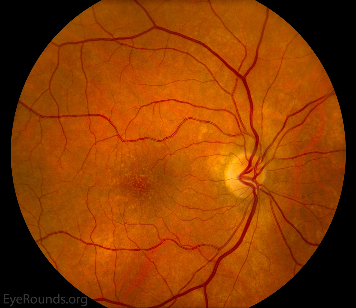
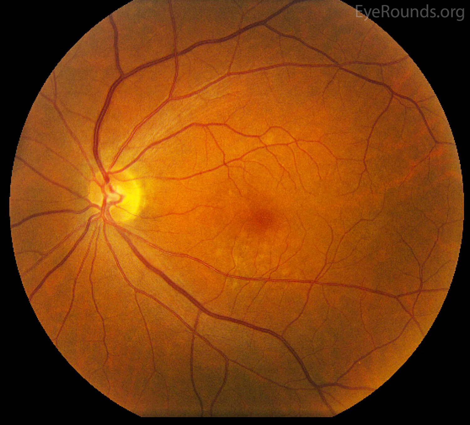
Early AMD (AREDS Category 2)
Figure 1a. These fundus photos show early AMD of two different patients. There are a few (<15) intermediate-sized soft drusen predominantly in the inferior macula.
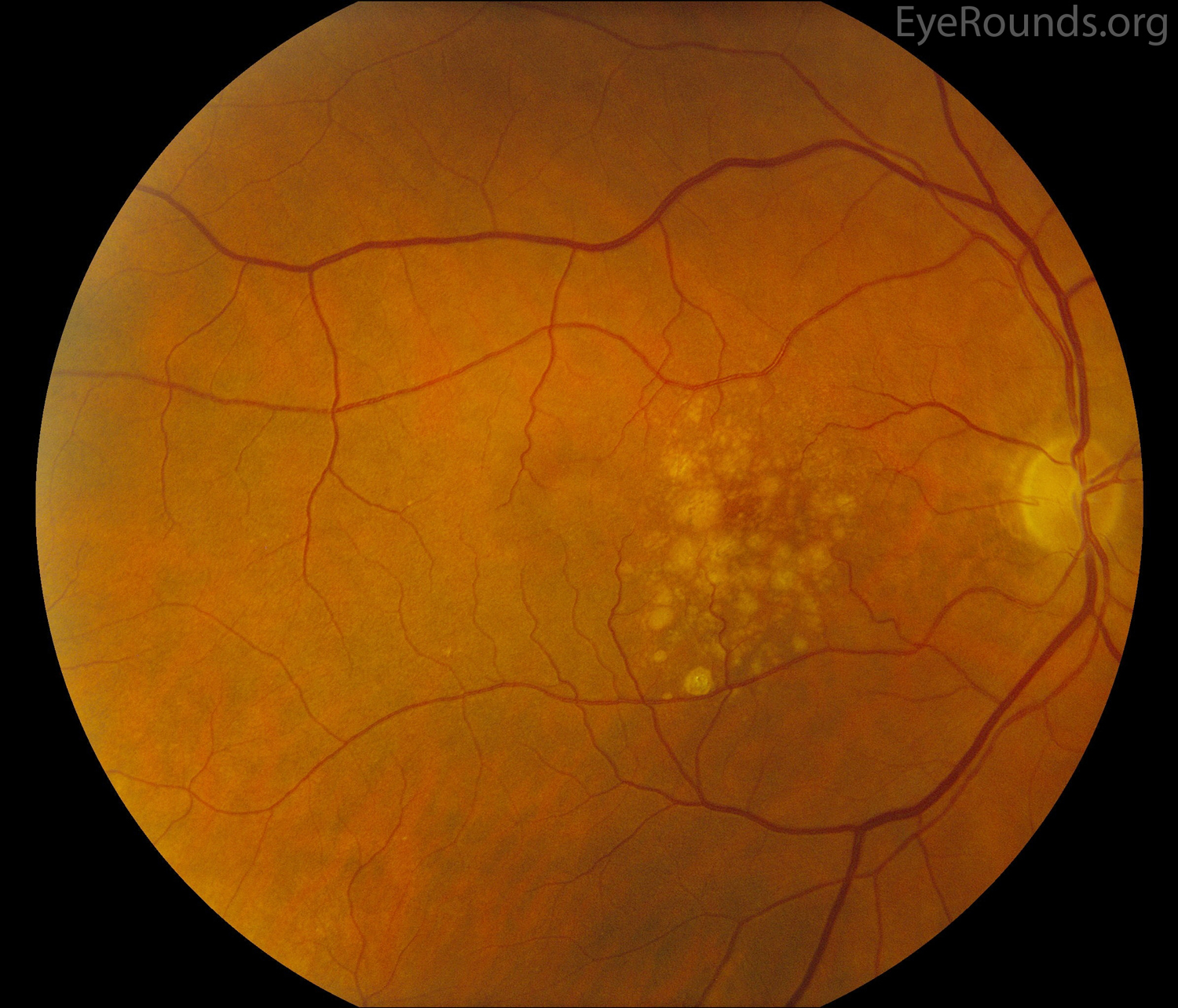
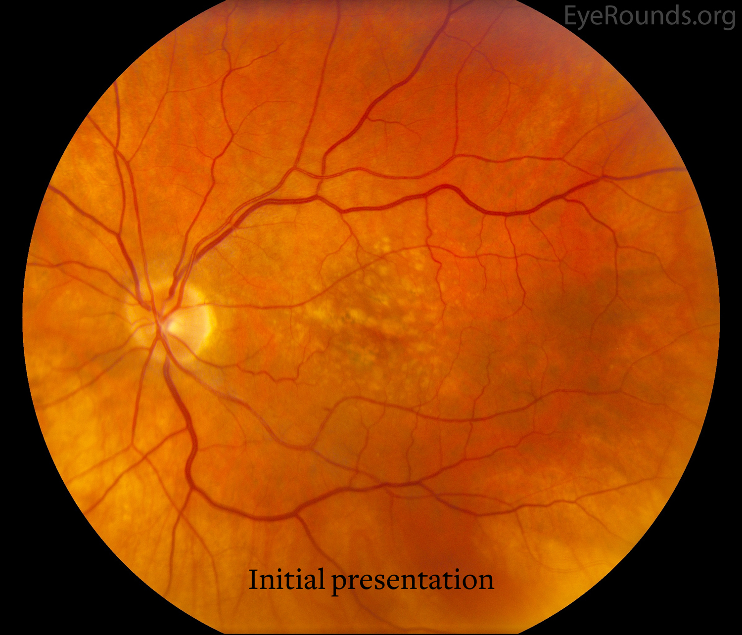
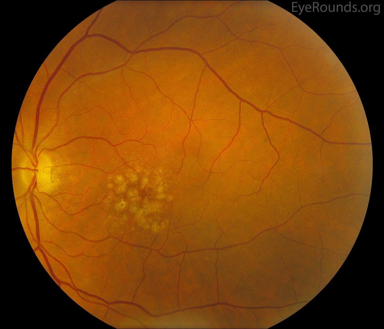
Figure 2a. Eyes demonstrates intermediate AMD with high risk (i.e., large, confluent, soft drusen) non-neovascular, age-related macular degeneration. Ultimately, the patient progressed to category 4 AMD with development of a choroidal neovascular membrane in the left eye (not shown), which required serial intravitreal injections.
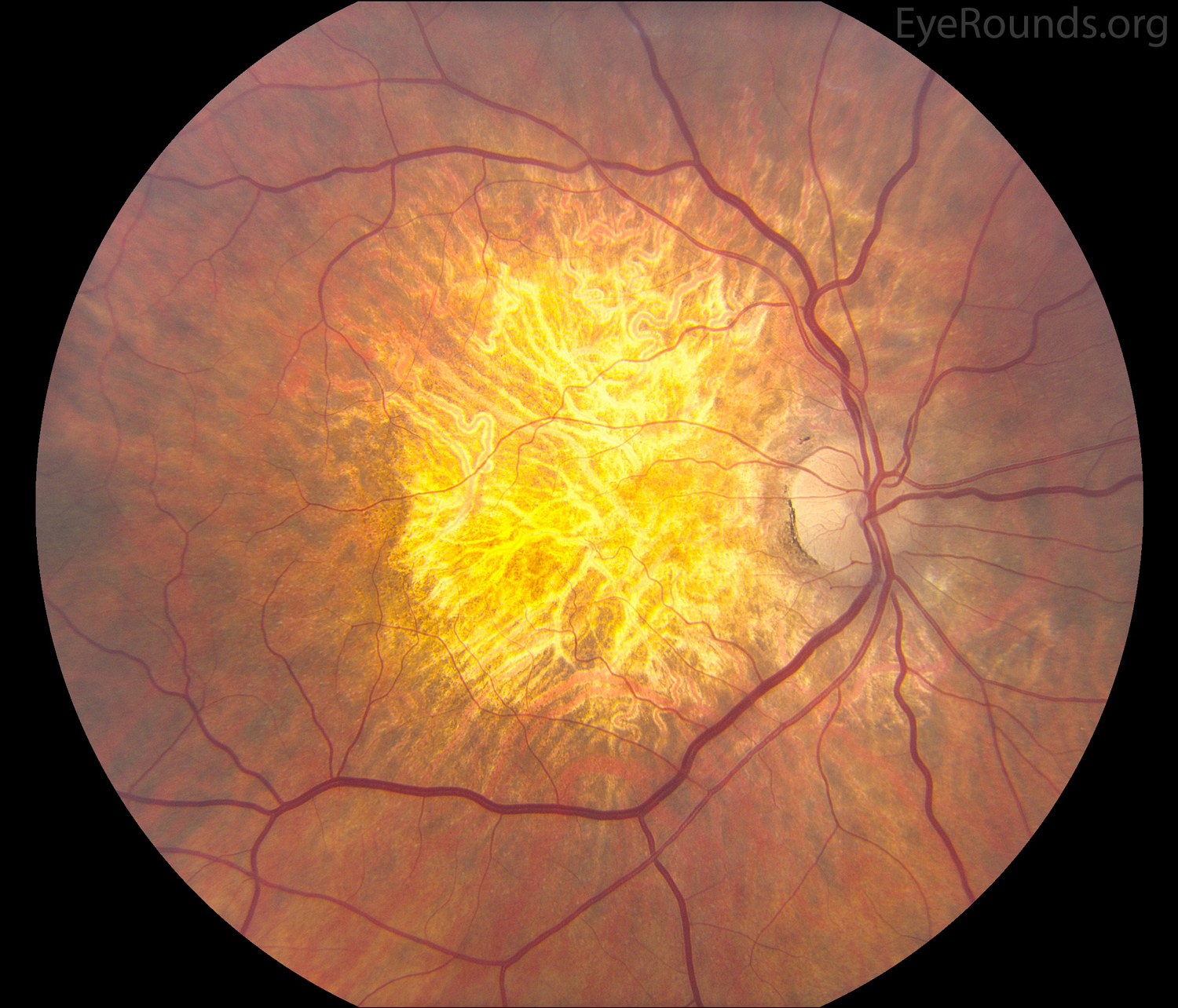
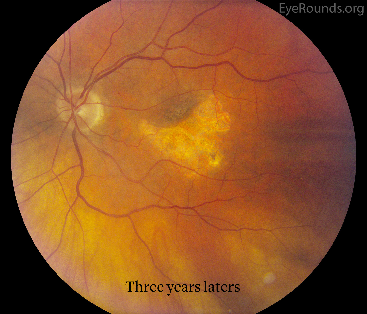
Figure 3b. Three years later, there was evidence of geographic atrophy only in the left eye, and the patient had significant vision loss in the left eye.
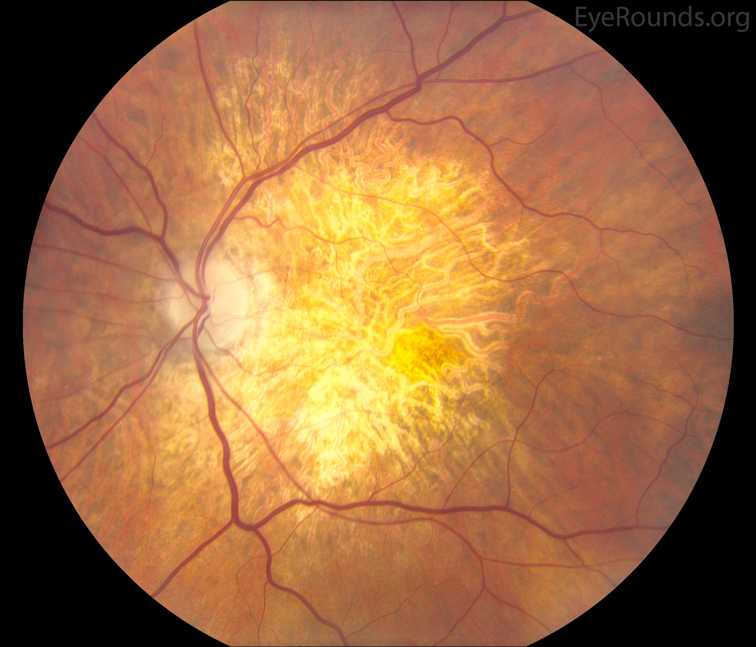
Figure 4a. Non-exudative macular degeneration with extensive geographic atrophy in both eyes. Since this patient had advanced AMD in both eyes, he was not an AREDS vitamins candidate.
Figure 4b. Non-exudative macular degeneration with extensive geographic atrophy in both eyes. Since this patient had advanced AMD in both eyes, he was not an AREDS vitamins candidate.
Wong WL, Su X, Li X, Cheung CM, Klein R, Cheng CY, Wong TY. Global prevalence of age-related macular degeneration and disease burden projection for 2020 and 2040: a systematic review and meta-analysis. Lancet Glob Health 2014;2(2):e106-116. https://PubMed.gov/25104651. DOI: 10.1016/S2214-109X(13)70145-1
Gehrs KM, Anderson DH, Johnson LV, Hageman GS. Age-related macular degeneration--emerging pathogenetic and therapeutic concepts. Ann Med 2006;38(7):450-471. https://PubMed.gov/17101537. DOI: 10.1080/07853890600946724
Group A-REDSR. The Age-Related Eye Disease Study (AREDS): design implications. AREDS report no. 1. Control Clin Trials 1999;20(6):573-600. https://PubMed.gov/10588299
Group A-REDSR. A randomized, placebo-controlled, clinical trial of high-dose supplementation with vitamins C and E, beta carotene, and zinc for age-related macular degeneration and vision loss: AREDS report no. 8. Arch Ophthalmol 2001;119(10):1417-1436. https://PubMed.gov/11594942
Woodell A, Rohrer B. A mechanistic review of cigarette smoke and age-related macular degeneration. Adv Exp Med Biol 2014;801:301-307. https://PubMed.gov/24664711. DOI: 10.1007/978-1-4614-3209-8_38
University of Iowa
Roy J. and Lucille A. Carver College of Medicine
Department of Ophthalmology and Visual Sciences
200 Hawkins Drive
Iowa City, IA 52242
University of Iowa
Roy J. and Lucille A. Carver College of Medicine
Department of Ophthalmology and Visual Sciences
200 Hawkins Drive
Iowa City, IA 52242