This site uses tracking information. Visit our privacy policy. Click to agree to this policy and not see this again.

Ophthalmology and Visual Sciences

Branched Retinal Vein Occlusion (BRVO)

Branched Retinal Vein Occlusion (BRVO)

Category(ies): Retina
Contributor: Christopher C. Robinson, MD and Jordan M. Graff, MD
Posted: February 8, 2008

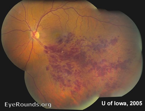
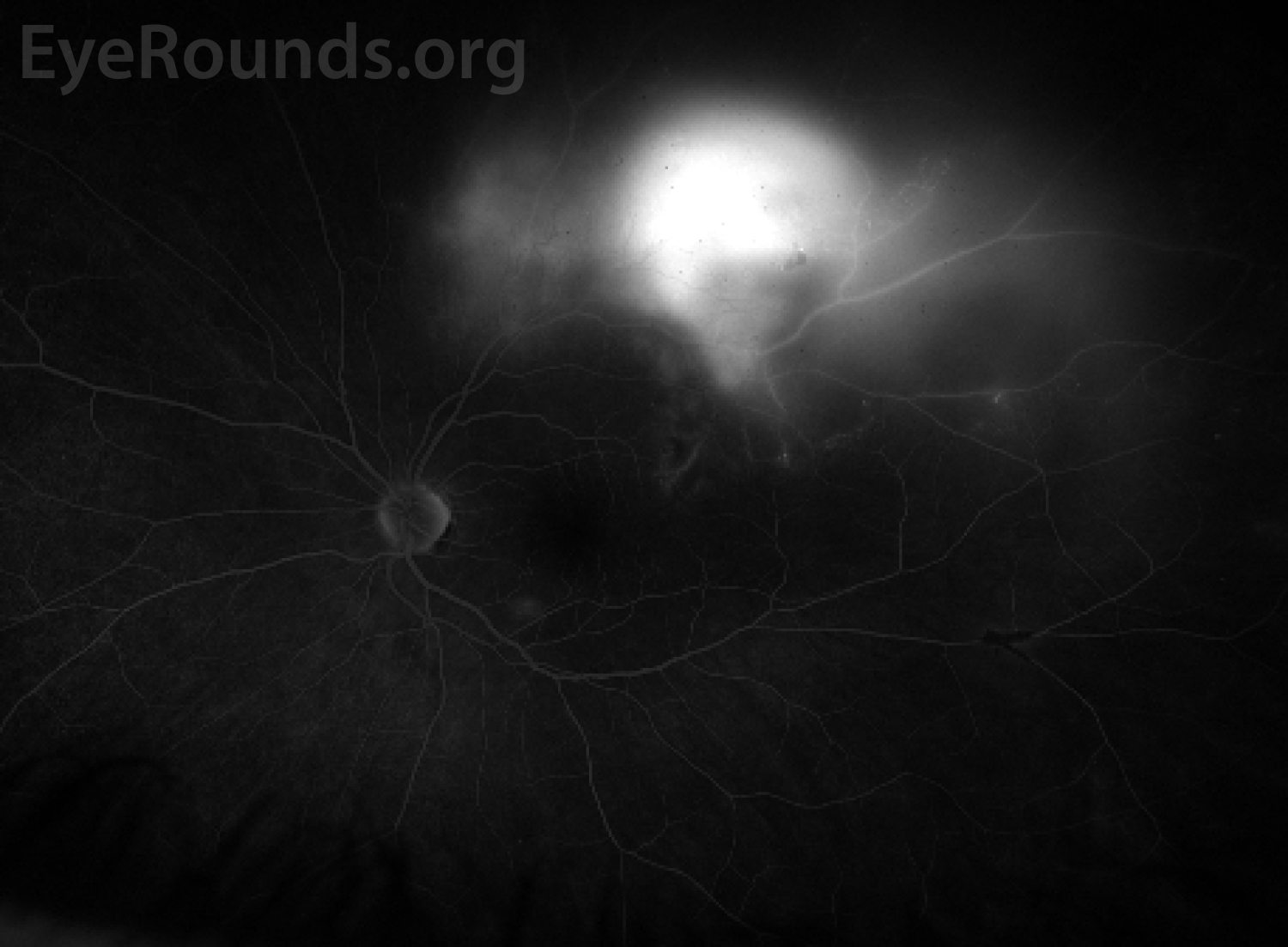
Classic sectoral hemorrhages are well-visualized in this case of Branched Retinal Vein Occlusion (BRVO). This condition is commonly associated with hypertension.


BRVO (Branched Retinal Vein Occlusion)

BRVO (Branched Retinal Vein Occlusion)

Category(ies): Retina
Contributor: Andrew Goldstein, MD and Michael D. Abramoff, MD, PhD
Contributor: Sarah Skiles, CRA
Posted: April 7, 2022

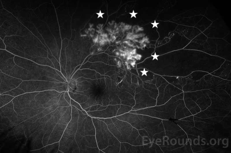
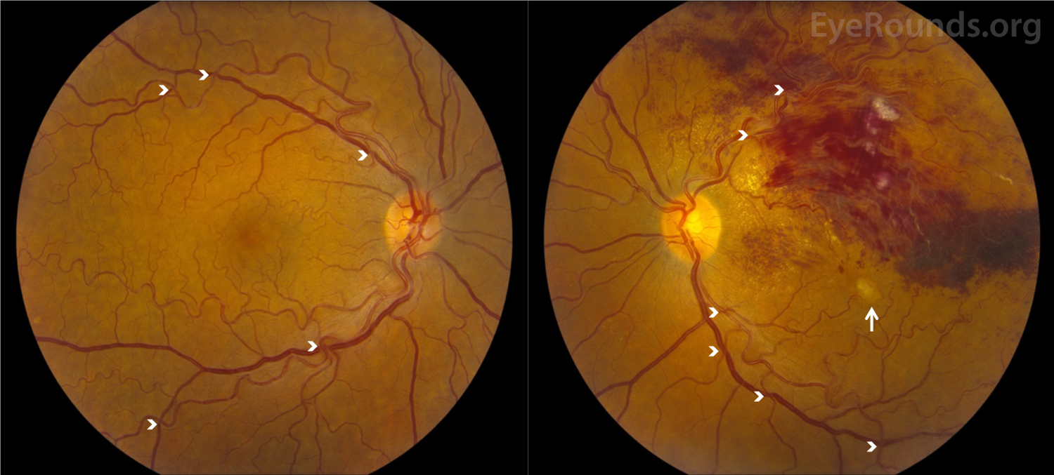
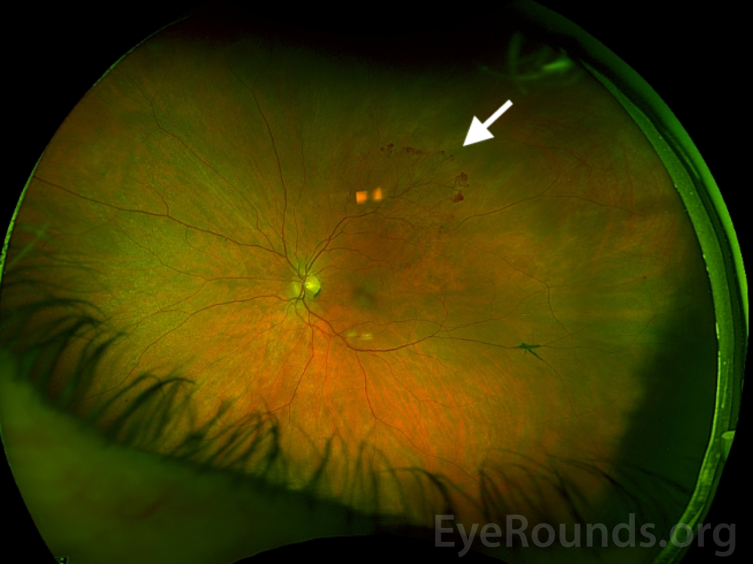
This series of images was obtained in a 69-year-old man who presented for a routine eye examination and reported a remote history of branch retinal vein occlusion (BRVO). He was asymptomatic with an uncorrected distance visual acuity of 20/20 in both eyes. On his initial examination he was noted to have a large area of peripheral neovascularization elsewhere (NVE). Scatter laser photocoagulation was applied to the large area of capillary non-perfusion anterior to the NVE in order to reduce the risk of hemorrhage.

Optos widefield imaging  superior temporal retinal neovascularization elsewhere
Figure 1: Optos widefield imaging of the left eye demonstrates superior temporal retinal neovascularization elsewhere (NVE) as indicated by the white arrow.
Optos widefield fluorescein angiography during the arteriovenous phase demonstrates neovascularization with mild leakage
Figure 2: Optos widefield fluorescein angiography during the arteriovenous phase demonstrates neovascularization with mild leakage and capillary non-perfusion of the surrounding retina (marked by white stars).
Optos widefield fluorescein angiography during the late phase shows continued superior temporal hyperfluorescence due to leakage from the neovascular vessels
Figure 3: Optos widefield fluorescein angiography during the late phase shows continued superior temporal hyperfluorescence due to leakage from the neovascular vessels.