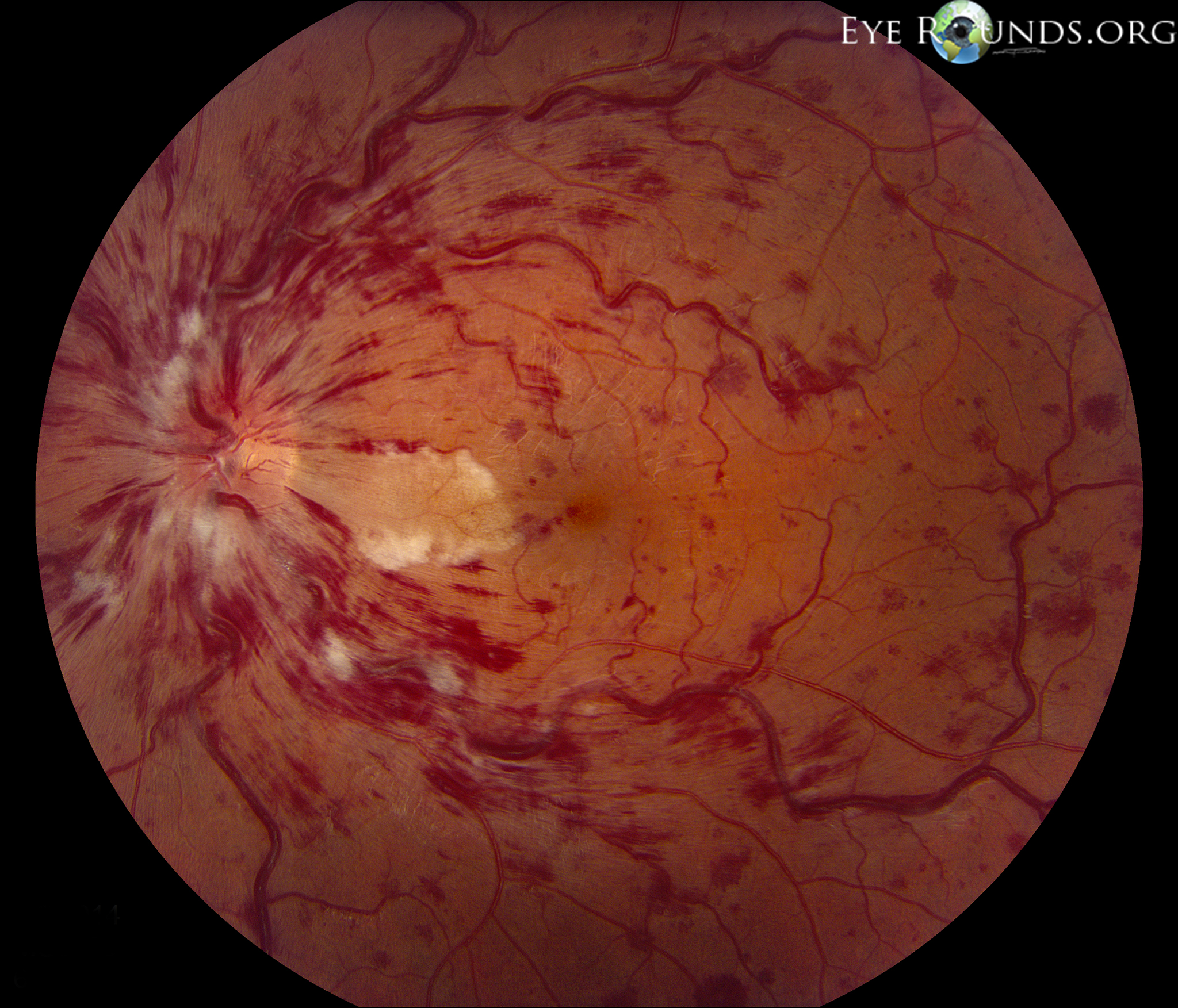
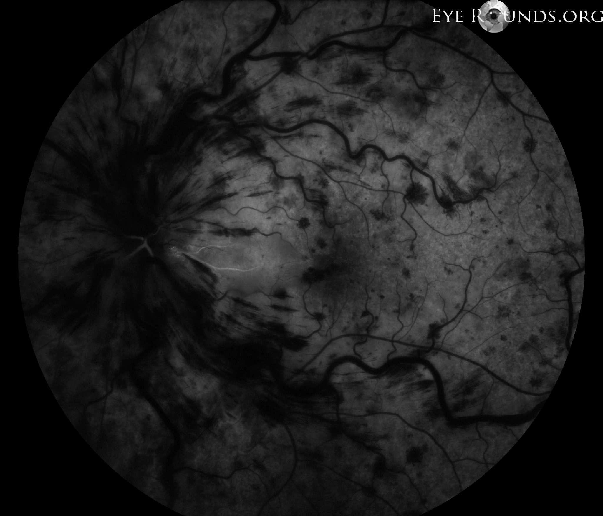
Cilioretinal artery occlusion (CLRAO) often occurs in the setting of a central retinal vein occlusion (CRVO). This lack of perfusion in the cilioretinal artery results from a functional hemodynamic block as the low perfusion pressure of the choroid, which supplies the cilioretinal artery, is unable to overcome the elevated venous pressure which results from the CRVO. The central retinal artery, however, is still able to perfuse due to its baseline higher perfusion pressure and ability to autoregulate its intraluminal pressure. In this photo, there are diffusely scattered intraretinal hemorrhages and cotton wool spots with associated venous distention and tortuosity secondary to the CRVO. Note the area of retinal whitening extending from the temporal disc into the macula in the region perfused by the cilioretinal artery. The fluorescein angiogram confirms that this is a cilioretinal artery as it fills with the choroid, prior to the filling of the central retinal artery.

Ophthalmic Atlas Images by EyeRounds.org, The University of Iowa are licensed under a Creative Commons Attribution-NonCommercial-NoDerivs 3.0 Unported License.

Address
University of IowaLegal
Related Links