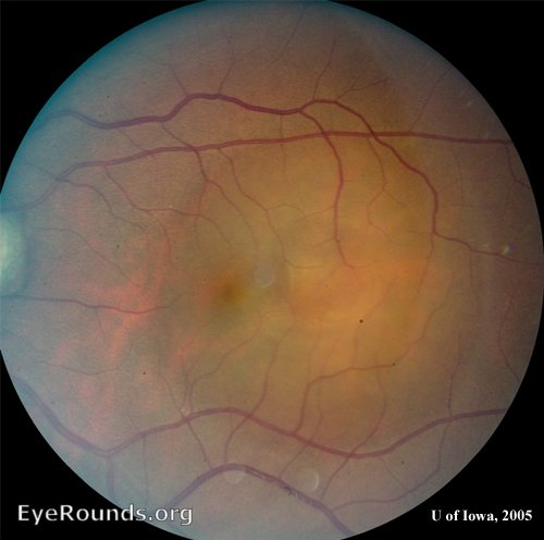
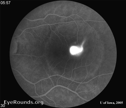
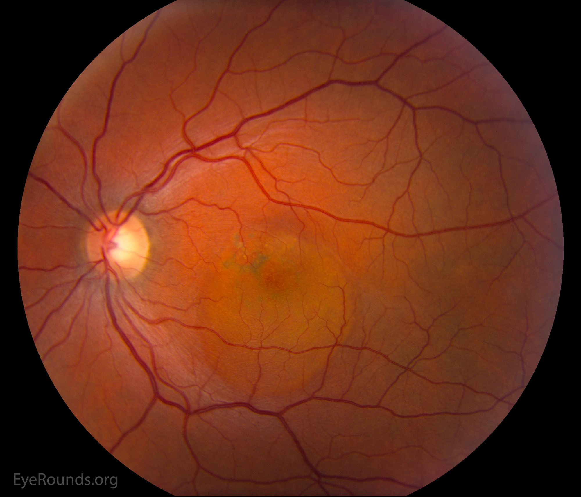
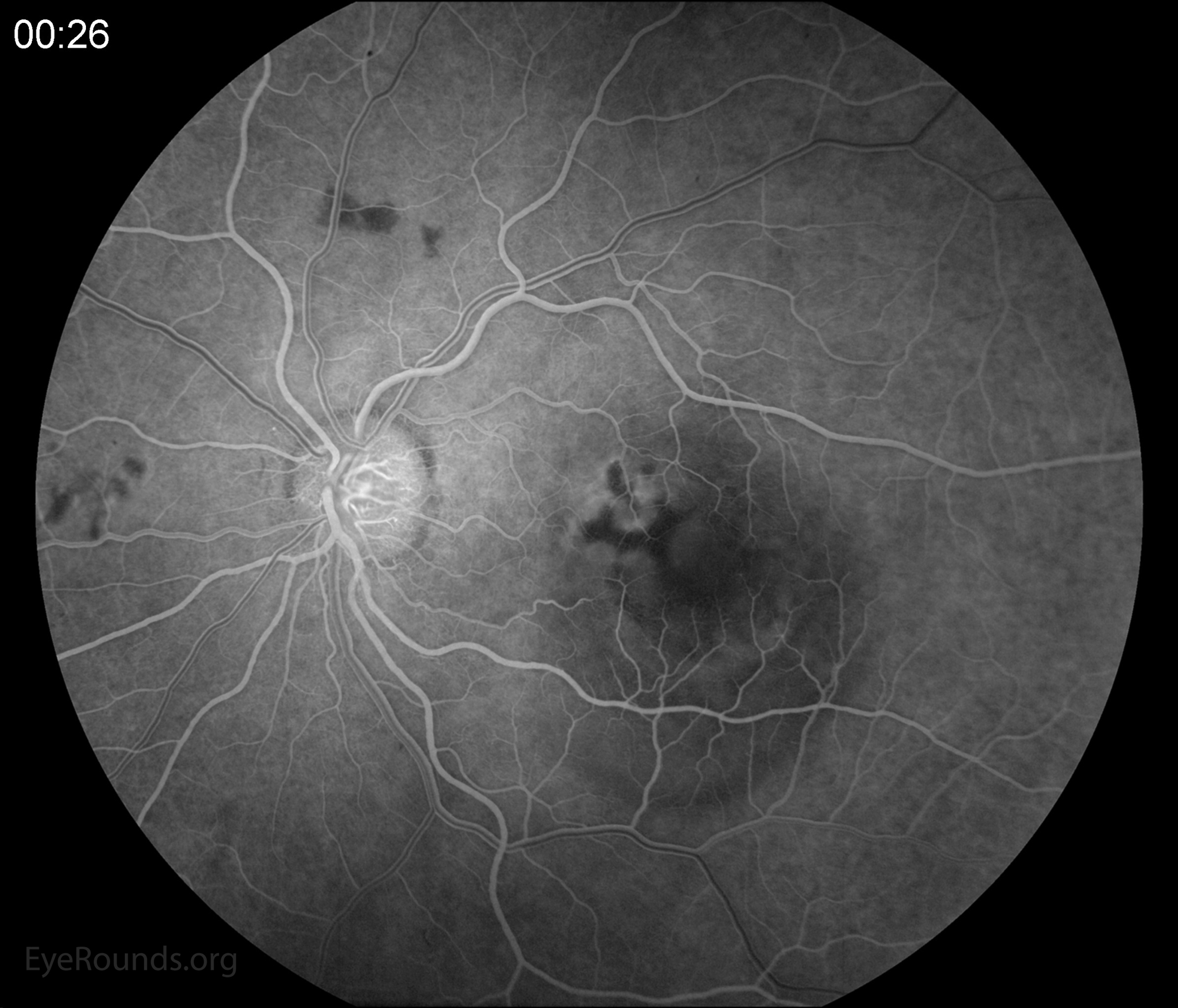
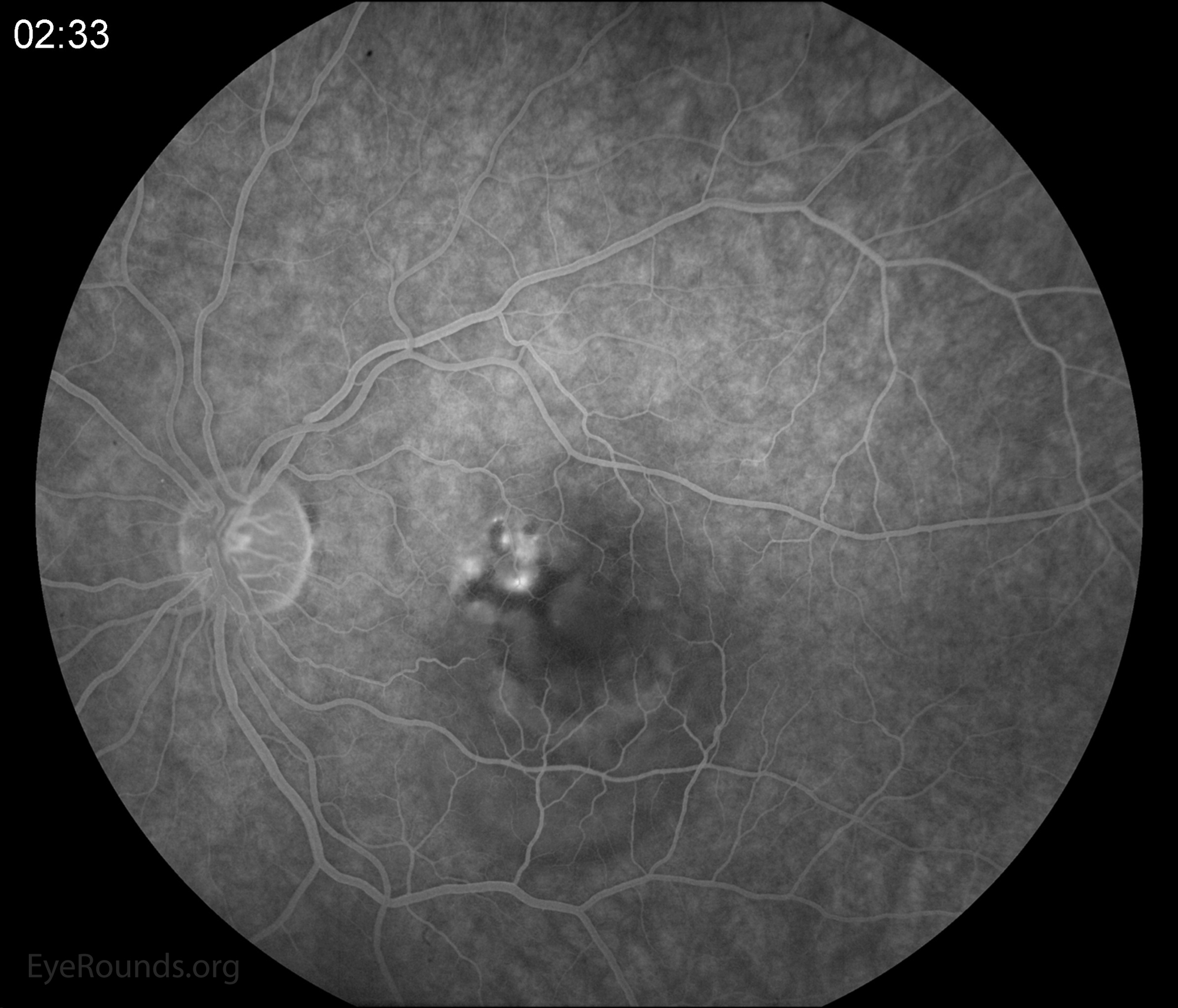
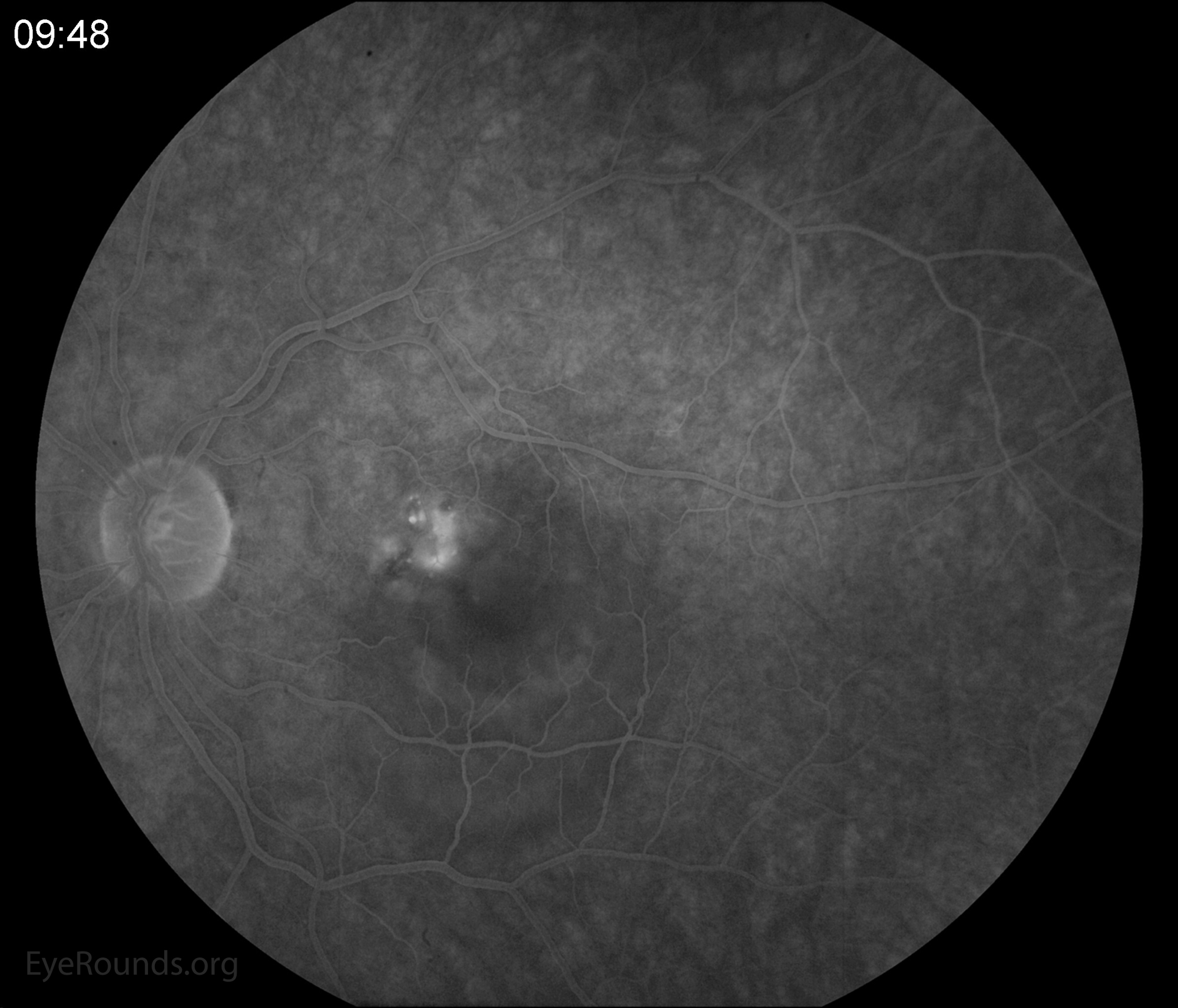
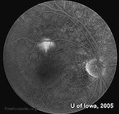
This color fundus photograph demonstrates subretinal fluid involving the fovea. There are multiple leakage points including the fovea on fluorescein angiography. In the late phase, one can see the classic "smokestack" leakage of dye.
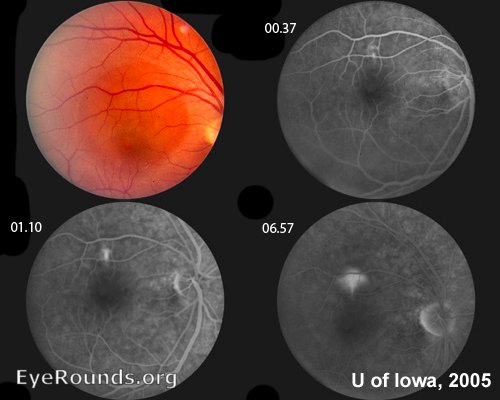
Color fundus photograph demonstrates subretinal fluid involving the fovea
Contributor: Jordan M. Graff, MD and Chet B. Patel, MD,
CSR (sometimes called Central Serous Chorioretinopathy or CSCR) is seen most commonly in male & female patients between the ages of 20 and 40 years of age.
Figure 1, a large, elevated area of serous retinal detachment can be seen occupying much of the temporal macula.
Within the central area of this detachment, there appears to be a second, smaller, ring of elevation. Flourescein angiography and optical coherence tomography (OCT) confirmed that this smaller elevated ring is a pigment epithelial detachment (PED) within the larger area of serous retinal detachment (see Figures 2-4 and discussion below).
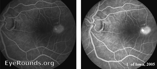
Early views of the flourescein angiogram demonstrate a hot spot of hyperflourescence that spreads to fill the PED. (In other classic cases of CSR, the hot spot may appear as a "smoke stack").
Late views with flourescein reveal the pooling of fluid within the serous detachement, correlating with the clinical appearance of the fundus in Figure 1.
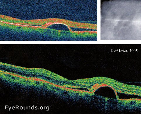
OCT images demonstrate a discrete blister of fluid underneath the RPE just temporal to the foveal depression. This defines the PED. The overlying serous retinal detachement is also evident.
A classic "smokestack" of leakage from the choroid is seen is this fluorescein angiogram. The fluid leaks into the subretinal space causing decreased visual acuity. Central serous retinopathy (CSR) is typically found in young males. There is a documented association with stress and steroid use.
University of Iowa
Roy J. and Lucille A. Carver College of Medicine
Department of Ophthalmology and Visual Sciences
200 Hawkins Drive
Iowa City, IA 52242
University of Iowa
Roy J. and Lucille A. Carver College of Medicine
Department of Ophthalmology and Visual Sciences
200 Hawkins Drive
Iowa City, IA 52242