

EyeRounds Online Atlas of Ophthalmology
Contributor: Matt Ward, MD, The University of Iowa
Category: Cornea
DMAEK (Descemet's Membrane Automated Endothelial Keratoplasty)
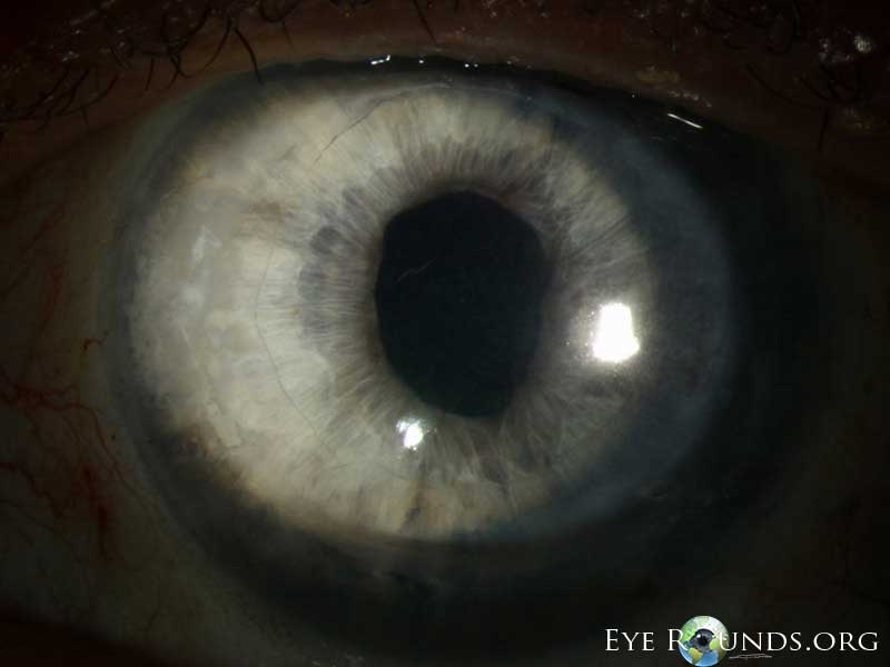
Image Comments:
1 month postoperative appearance. Note stromal donut with clear central Descemet's membrane graft.
DMAEK is a hybrid of DMEK and DSAEK. The Iowa Lions Eye Bank prepares the graft using a lamellar Femtosecond laser cut deep into the stroma and then uses air dissection (much like in DALK surgery) to cleave a pre-Descemet's plane in the center of the graft (usually about 6 mm in diameter). The tissue is then placed back in Optisol GS media and packaged for the surgeon. In the OR the surgeon trephinates the donor usually at 8.0 or 8.5 mm so that the central Descemet's membrane is surrounded by a rim of stroma (approximately 120 microns thick). The stromal ring allows for easier manipulation within the eye (much like DSAEK), but the central Descemet's membrane graft allows for speedy clearing and likely improved end visual acuity (though rigorous study of the results of DMAEK have yet to be completed).
No one may use these photographs for financial gain without written authorization from the contributor.

Ophthalmic Atlas Images by EyeRounds.org, The University of Iowa are licensed under a Creative Commons Attribution-NonCommercial-NoDerivs 3.0 Unported License.